
Story at a Glance
•DMSO is an “umbrella remedy” whose combination of therapeutic properties makes it uniquely suited to treat “incurable” conditions. This article will focus on the extensive evidence DMSO safely eliminates clots, protects the blood vessels, and restores circulation, as impaired blood flow is one of the most common causes of disease but challenging to manage with conventional options.
•DMSO revolutionizes the treatment of strokes, as it not only eliminates the stroke, but also can be started hours earlier than normal treatments and is able to heal the injured tissue that would otherwise be lost. Extensive evidence, and now reader reports show that were it to be widely adopted, millions could be spared from the disability or death they cause each year.
•DMSO is also uniquely suited to treating brain bleeds, traumatic brain injuries, and concussions, as it relieves pressure on the brain while restoring vital circulation and reviving dying brain cells. Extensive evidence and reader reports again show its utility here.
•Because the heart, like the brain, is exquisitely sensitive to its blood supply being cut off, many of DMSO’s properties have also been repeatedly shown to have immense value in heart attacks alongside many other severe heart conditions.
•DMSO has been extensively used for peripheral circulatory disorders, with hundreds of readers, mirroring the existing evidence, reporting it eliminating varicose veins present for decades, dissolving clots and DVTs, restoring color and sensation to blue, purple, or black ischemic limbs, healing venous, diabetic, and pressure ulcers, and resolving Raynaud’s, frostbite, and hemorrhoids, frequently within days.
•This article summarizes the extensive data demonstrating DMSO’s efficacy for circulatory and vascular conditions (approximately 1500 studies and 160 pertinent reader testimonials) and provides practical guidance on DMSO protocols and complementary approaches for the neurological and vascular conditions discussed throughout this series.
Decades ago, I learned that DMSO was an excellent therapy for strokes and then subsequently had numerous experiences where someone I told to take DMSO had their full stroke resolve on the way to the hospital (sparing them from a life of expected disability), after which I started also using DMSO for successful post-stroke recoveries. When you consider both how debilitating strokes typically are and that, for decades, there has been only piecemeal progress on reducing the suffering they cause, it’s hard to put into words how upsetting this has been for me.
So, once I realized I’d stumbled upon being the custodian of a widely read publication, one of my first major projects was to write “The Evidence DMSO Could Save Millions From Brain and Spinal Injury,” which struck the same chord in many readers that it had in me, and was seen by millions (many of whom subsequently shared DMSO saved them from a stroke).
In the process of researching that article and the reader requested ones which followed (on the myriad of other transformational uses for DMSO), I gradually realized that a massive volume of buried DMSO literature existed, and felt that due to the support I’d received from readership here, I had the obligation to make that lost knowledge (and the thousands of reader DMSO testimonials—currently 7,500) widely available.
Note: some of the most dramatic neurological recoveries readers shared (which can be viewed in filmed interviews) include from terminal ALS, terminal bulbar ALS, advanced CJD (the 100% fatal prion disease), sight that had been lost for nearly 75 years, and the same spinal cord injury that left Christopher Reeve on a vent for life.
The fastest way I could see to actually do that was to search the key English and foreign databases for each term for DMSO (rather than every possible keyword combination), then filter out the medically relevant studies and compile them and condense them into articles.

Given how much was there, I was initially unsure if this was possible, but over six grueling months (where I was forced to cut back on everything else and hence published less here), I learned a lot, and with the tools that have never before been available, eventually collected 15,000 summarized studies.
Note: in the process of doing the same for the medical ozone literature base, I recently discovered there is one large DMSO database I missed that will need to be done in the near future.
Once they were compiled, I polled the supporters here on which topics had the most interest, and the overwhelming answer was neurology. On one hand I supported starting there, as most of the therapeutic mechanisms which underlie how DMSO works are interwoven with neurology (and I believe laying out the mechanistic basis for how DMSO works is critical for the therapy’s adoption), but I was also hesitant as this was single most challenging part of the topic to synthesize (due to the ultimately 4,500 studies which would need to be incorporated).
Nonetheless, starting at the end of April, I was able to begin publishing them, and this article, at last, marks the final part of that series. The previous ones, which provided the mechanistic basis for much of what is discussed in this article are as follows:
How DMSO Heals the Brain and Transforms Neurology
This article covers how circulatory impairments underlie many neuropsychiatric disorders and how DMSO:
•Biophysically eliminates microclots along with preventing immune cells from obstructing capillaries, facilitating lymphatic and venous drainage.
•Counteracts a broad range of inflammation and free radicals
•Shields cells from diverse lethal stressors and excitotoxins
•Protects ATP production, restarts dormant cells, and restores parasympathetic (vagal) tone (e.g., through acetylcholinesterase inhibition)
•Effectively delivers a variety of therapeutic agents to the central nervous system.
How DMSO Heals the Spine and Reverses Paralysis
This article also extensively covers:
•How DMSO facilitates neuronal regeneration by stabilizing microtubules and differentiating stem cells into replacement neurons.
•How DMSO’s other established therapeutic mechanisms (e.g., muscle relaxation, tissue regeneration, and interruption of pain transmission) reduce or eliminate spinal pain.
How DMSO Heals The Nerves & Eliminates Pain

This article also extensively covers how:
•DMSO’s biophysical effects on water temporarily shift the phase of cell membranes and the cytoskeleton, creating a reversible cellular reset that restores normal structure and function (along with uniquely enabling DMSO to transport substances across biological barriers without damaging them).
•DMSO selectively blocks the small nerve fibers that transmit chronic pain
•DMSO supports peripheral nerve regeneration (e.g., with membrane resealing)
•Through neuronal resets, DMSO can eliminate the source of chronic pain and autonomic dysfunction.
In this article, I will cover one of DMSO most extensively studied mechanisms: how it works through several well-established anticoagulation pathways to reduce clotting, dilate vessels, protect blood vessels and improve circulation (which works synergistically with its biophysical ability to eliminate micro-clotting discussed in the first part of this series).
Note: if the information in the next section is too dense, you can skip ahead to the “Current Stroke Management” section.
Effects on Platelets
Platelets, the tiny cell fragments that clump together to initiate clotting, are one of the most common targets of blood-thinning medicines (e.g., aspirin) and likewise one of the most thoroughly studied targets of DMSO. Collectively, this evidence shows DMSO independently inhibits platelet aggregation, adhesion, and activation, and most importantly, does so through several converging mechanisms at once.
For example, one study examining the full panel of platelet agonists (the natural triggers that switch platelets on) found DMSO inhibited human platelet aggregation induced by ADP, adrenaline, arachidonic acid, collagen, and PAF, with the strongest effect against ADP. Multiple independent groups, in turn, have reproduced these results.1,2,3
Likewise, across studies, DMSO has been found to inhibit platelet aggregation (clumping) triggered by ADP, epinephrine (adrenaline), arachidonic acid, collagen, thrombin, and platelet-activating factor, in a concentration-dependent manner, with ADP-induced aggregation typically the most sensitive to DMSO.1,2,3 At the level of activation markers, DMSO-containing systems suppress the surface proteins that report a platelet has “switched on”—P-selectin (CD62P), PAC-1, GPIIb/IIIa, CD63, and platelet factor 4 (PF4)—and reduce the release of ATP and dense-granule contents.1 DMSO also directly binds platelet factor 4, disrupting its aggregation at the molecular level.
Note: these effects are concentration and time dependent. At the low-to-moderate concentrations relevant to therapeutic use, the antiplatelet effect reverses once DMSO is washed out—consistent with DMSO's rapid diffusion out of cells, which is why platelets cryopreserved in DMSO lose adhesion and aggregation during cold storage yet regain functionality once thawed and infused.1,2 At the higher concentrations or longer exposures reached only outside normal use, the inhibition can persist even after removal,1 reflecting a more durable structural change. However at the higher doses, such as the concentrations reached after a DMSO-cryopreserved cell infusion (0.2–0.6%), DMSO still measurably inhibits ADP, thrombin, and TRAP-induced platelet aggregation,1,2 frequently causes transient circulating coagulation inhibitors and prolonged clotting times1,2 and at high topical doses can modestly lower the circulating platelet count.1
•DMSO selectively inhibits cyclooxygenase-1 (COX-1) and thromboxane A₂ synthase,1,2 (e.g., 0.5% DMSO reduced total COX activity by 36%1) cutting production of thromboxane A₂ (the potent signal platelets use to recruit one another). Adding the synthetic thromboxane analogue U46619 restored platelet aggregation despite DMSO’s presence,1 confirming that DMSO’s suppression of aggregation runs through the thromboxane pathway.
•DMSO raises intracellular cAMP and cGMP (the secondary messengers that hold platelets in the resting state) providing a thromboxane-independent brake on platelet activation.
•Because DMSO restructures the water in and around the cell membrane (detailed here), it alters membrane fluidity directly, desensitizing platelets to their agonists independent of COX-1—an effect visible under the microscope as a marked reduction in the filopodia platelets extend when activating.1 This biophysical membrane action also likely explains DMSO’s suppression of shear-induced platelet activation,1 where DMSO cut platelet adhesion under flow to roughly a fifth of control,1 and connects to the same phase-transition effects DMSO exerts on the platelet membrane’s phospholipids.1 A separate study of platelets forced through the non-physiological shear stresses generated by mechanical heart-assist devices found the same protection, and in a revealing pattern that mirrors the concentration-dependent membrane stages described earlier: low concentrations of DMSO sharply reduced shear-induced activation, while the effect reversed at higher concentrations as DMSO began permeabilizing the membrane—and DMSO-treated platelets, though shielded from mechanical activation, still responded normally to chemical agonists, confirming it desensitizes platelets to physical force rather than simply disabling them.1 Notably, in that same body of work the conventional antiplatelet drugs, designed to block platelets through specific chemical pathways, were overwhelmed by these device-level physical forces, whereas DMSO, acting on the membrane itself, was not.1 At higher concentrations this progresses to increased membrane permeability, at which point DMSO releases the platelet’s internal contents through a mechanism distinct from thrombin’s.1
•DMSO reversibly inhibits platelet serotonin uptake, thereby (reversibly) reducing the amount of serotonin stored in (and later released from) dense granules that is available to amplify platelet aggregation.1
•Since hydroxyl radicals trigger platelet serotonin release and aggregation, DMSO’s extensively documented scavenging of these radicals1 blocks radical-mediated platelet activation1 and thrombus (clot) formation.1 In living microvessels, this was directly visualized: intraperitoneal DMSO more than doubled the time to platelet aggregation in injured mouse brain pial arterioles (117 vs. 57 seconds), and in the same vessels prevented the reactive arteriolar dilation that normally accompanies the injury (holding their diameter near baseline versus a 22% widening in controls).1 As such, DMSO’s radical-scavenging action, in addition to reducing inflammation, also produces both an antiplatelet and a vasomotor effect simultaneously.
Note: as each of these effects is concentration dependent, not every study finds an effect on every pathway (e.g., one group reported DMSO did not inhibit thromboxane production in platelets and did not protect rabbits from arachidonic-acid–induced pulmonary thrombosis1,2).
These effects, in turn have been demonstrated their utility in a variety of living systems:
•In a dog model of critical coronary stenosis (where platelets aggregating at a narrowed artery routinely cause cyclic drops in coronary blood flow) intravenous DMSO dose-dependently reduced (by up to 85%) those flow reductions without altering blood pressure, heart rate, or cardiac output, isolating the effect to platelet aggregation itself.1
•In a rat carotid-injury model, IV DMSO (2 g/kg) virtually abolished thrombus formation despite clear endothelial damage, with electron microscopy showing only spherical, non-activated platelets where clots should have been.1,2
Note: because the clotting was prevented at a site of genuine vessel injury, these results1 are applicable to settings like microvascular and reattachment surgery, where a damaged vessel must stay patent long enough to heal.
Finally, DMSO’s effects on platelets (inhibition of aggregation and suppression of activation markers such as P-selectin, PAC-1, GPIIb/IIIa, and PF4) have also been observed when DMSO has been combined with a variety of natural compounds (which for reader ease I mark with ⬖) such as xanthohumol,⬖ wine polyphenols,⬖ Ginkgolide B,⬖ and vitexicarpin.⬖
One of the more unusual reports came from a Russian practitioner combining DMSO with aspirin in "structured water," across a range from conventional doses down to ultra-low dilutions. Using nocturnal leg cramps in the elderly as a readout, he reported that conventional doses reliably prevented recurrence — cramps returned on withdrawal and stopped again on resumption — with the effect persisting even as aspirin was diluted roughly a thousandfold, becoming borderline around the nanogram range (~10⁻⁹) and disappearing by ~10⁻¹². He also described rapid relief in acute vascular events such as heart attack and stroke.1
Anticoagulant & Fibrinolytic Effects
DMSO also acts directly on the coagulation cascade itself, and does so at nearly every level (the initiating trigger, the central enzyme thrombin, and the fibrinolytic system that dissolves clots once they form) giving DMSO a breadth of anticlotting activity that few single agents possess:
•Tissue factor (TF) is the key protein that ignites the entire clotting cascade (and is a critical link between inflammation and thrombosis). DMSO, in turn, suppresses TF expression and activity by inhibiting JNK and p38 MAP kinase signaling and prevents arterial thrombus formation in living animals, without detectable toxicity.1,2 Because it simultaneously reduces the smooth-muscle overgrowth that causes stents to re-occlude, DMSO has been repeatedly proposed as a drug-eluting stent coating due to it being a rare agent that both lowers clotting risk and restenosis risk (unlike conventional stent drugs like sirolimus and paclitaxel, which reduce restenosis but increase TF expression and thrombosis risk).1,2,3
•DMSO also acts on the coagulation enzymes themselves. It directly alters thrombin, the central clotting enzyme, changing its catalytic behavior through a conformational shift and—notably—reducing heparin’s ability to inhibit it.1 In the invertebrate clotting systems used as sensitive models of the cascade, DMSO reversibly blocked the activation step that switches on the dormant clotting zymogen (enzyme precursor), without inhibiting the already-active enzyme, in both the Limulus and Carcinoscorpius systems (highly sensitive clotting cascades)1,2—indicating DMSO acts on the trigger that starts the clotting cascade.
DMSO hence not only prevents clots from forming but, when applied topically, actively dissolves already-established venous thrombi while inhibiting arterial platelet thrombus formation. Finally, in a rat carotid-artery injury model intravenous DMSO eliminated thrombus formation at both crush and incision sites despite clear endothelial damage. Electron microscopy showed only minimal platelet-fibrin deposits and, spherical, non-activated platelets where clots should have formed (with DMSO’s antithrombotic effect statistically superior to the barbiturates and methylprednisolone tested alongside it).
Its ability to eliminate clots already present appears to be due to it augmenting the body’s natural clot removal pathways as DMSO activates streptokinase-initiated fibrinolysis across a wide concentration range (1.5–50%), stimulates production of tissue plasminogen activator (tPA), the body’s principal clot-dissolving enzyme and reduces plasminogen activator inhibitor-1 (PAI-1), the main physiological brake on that process.
This is cited so consistently that “fibrinolytic activity” appears as a standard listed property of DMSO throughout the clinical literature,1,2,3,4 or as described by a Russian review: intravenous DMSO lengthens clotting time, suppresses platelet aggregation (initial clot formation), normalizes fibrin formation (clot solidification), and being fibrinolytic, normalizes fibrinolysis.1
All of this touches on a key contrast I’ve noticed between pharmaceutical drugs and effective natural therapies. Pharmaceuticals tend to target a single pathway (or a few) as forcefully as possible, which produces both their immediately evident effects and, frequently, significant side effects. Time-tested natural agents instead often nudge many pathways at once toward a converging end, making them less immediately potent (their effect being distributed rather than concentrated) but also much better tolerated and often longer-lasting, since the body doesn’t develop the reflexive tolerance it mounts against a pathway being aggressively shifted in one direction.
Note: as with many of DMSO’s effects, its fibrinolytic action normalizes rather than simply pushing one direction—in cancer cells it suppressed urokinase-type plasminogen activator (uPA—which facilitates metastasis), the reverse of its tPA-stimulating effect in normal cells, consistent with DMSO restoring dysregulated systems toward baseline.
Clinically, this translates to DMSO improving coagulation across a broad range of situations. For example, in 42 patients with rheumatic diseases (including scleroderma and Raynaud’s), blinded thromboelastography showed DMSO normalized both fibrin formation, with accompanying improvement in microcirculation. Likewise, DMSO added to sepsis, endotoxemia, and disseminated-intravascular-coagulation models and paired with a long and chemically unrelated list of agents, has repeatedly reduced elevated fibrinogen and corrected the underlying coagulopathy.1,2,3,4,5 Similarly, DMSO appears in numerous topical thrombosis and thrombophlebitis formulations where it both facilitates the delivery of and synergistically enhances the effects of the other anti-thrombotic agents present.1
Note: DMSO’s broad range of effects also allows it to treat challenging clotting situations. For example, when the immune system breaks down myelin, the exposed phospholipids in the debris can trigger clotting that starves nerves of blood (a proposed secondary driver of multiple sclerosis) and while conventional anticoagulants don’t touch this pathway, a Russian study found DMSO inhibited myelin-triggered clotting dose-dependently.
DMSO and Blood Thinners
In medicine a balance often has to be struck between a therapeutic effect and the toxicity it entails, and getting it right can be quite challenging, such as in ensuring chemotherapy kills enough cancer cells without killing too many normal ones. Blood thinning poses another version of this problem: coagulation can predispose someone to severe complications (e.g., embolic strokes), but too little of it is also dangerous, since a minor injury can then produce a large hematoma and a blow to the head a lethal brain bleed.
For this reason, medicine typically reserves anticoagulants for those already at risk of clotting complications, as in atrial fibrillation, where the irregular rhythm lets clots form in the heart that can then travel to the brain and cause a stroke. It then tightly controls the degree of anticoagulation, doing enough to prevent the major event without raising bleeding risk any more than necessary. However, this is still far from perfect. Many individuals experience significant complications from anticoagulants each year, partly from incorrect dosing or monitoring, and partly because a perfect balance between preventing thrombosis and avoiding bleeding simply cannot always be achieved in the individual patient. All of this hence raises a few questions regarding DMSO.
First, can natural “anticoagulants” with a broader spectrum of action be used in lieu of the more potentially dangerous anticoagulants? Ultimately this is a difficult question to answer, as understandably, no clinician I know has taken the risk to attempt this in high risk situations (e.g., atrial fibrillation) and see if it suffices to stop the clotting (whereas gargantuan sums of data were needed to accurately assess the merit of pharmaceutical anticoagulants). However, in lower risk situations (e.g., preventing a heart attack over the next twenty years, or improving chronic illness symptoms resulting from excessive blood clotting), I have seen the natural alternatives perform extremely well without significant risk. Likewise, in acute situations (e.g., a heart attack or stroke), I have seen numerous cases where they (e.g., DMSO or a zeta potential restoration) resolved the incident prior to conventional options arriving. A few readers, in turn, have shared stories like quitting a problematic lifelong Eliquis prescription, noting DMSO not only prevented clots from forming but also dissolved existing ones and that “I’d be dead if DMSO didn’t work.”1 I, however, do not think this is wise, as the data simply does not exist to determine if this is appropriate to do, and as such, I instead favor the natural anti-clotting approaches in cases where it seems likely someone would benefit from blood thinning but their inherent risk of a severe clotting complication is too low to justify conventional anticoagulation.
Note: while safer than conventional anticoagulants, many of the natural anticoagulants are still not completely safe (e.g., since mass consumption of generic nattokinase⬖ came into vogue to counteract the spike protein, I have periodically come across cases of them causing a problematic bleed both within my social circle and from readers here).
Second, since DMSO potentiates the actions of pharmaceutical medications, is it safe to take with other anticoagulants? Presently, there is simply not enough data to answer this, but from everything I’ve been able to find, I lean to “yes,” with three major caveats: DMSO cannot be taken within at least two hours of the anticoagulant, coagulation must be carefully monitored to ensure excessive anticoagulation is not occurring (which should happen anyways) and IV DMSO should never be mixed with an anticoagulant in the same infusion. The limited datapoints I have to go off for this assessment are that:
•The German DMSO community, which has the most experience combining DMSO with conventional anticoagulants, considers this acceptable, citing patient reports, existing data that DMSO does not competitively inhibit clotting factors or irreversibly block platelet prostaglandins the way pharmaceutical anticoagulants do, and that in practice no meaningful changes in Quick-Wert (PT activity %) or INR values appear in patients on anticoagulants who use DMSO—although they did note DMSO can reduce the dose of aspirin (which targets platelets rather than the coagulation cascade), with multiple users “switching to 50 mg every other day and their coagulation values still being ‘as desired.’” That said, they still advise starting DMSO slowly, routinely checking coagulation values, and spacing it out from anticoagulants taken during the day.
Note: in the previously mentioned Russian study (where very limited information was provided), DMSO was reported to work synergistically with aspirin and no toxicity was mentioned from this combination.1
•DMSO–heparin combinations have a long track record of safety in topical and oral formulations. DMSO enhances heparin’s anticoagulant action and, through its membrane-transport properties, can even render heparin orally absorbable when the two are formulated together (rather than via the injection typically required for heparin). Likewise, the commercial gel Dolobene (Долобене), which combined DMSO with heparin has been widely used for decades without issues (as has Phlebolan spray, although only certain Phlebolan formulations contained heparin).
Note: Jim McCann (the inventor who brought the DMSO hematoxylin cancer therapy to Ecuador) emphasized that he had seen serious bleeding issues when IV DMSO was combined with IV heparin.
•Due to the size of the readership of this newsletter, I've had a unique window for identifying rare complications of DMSO, so over the last two years, I've kept a careful eye out for bleeding incidents linked to DMSO, of which a few reported nosebleeds after DMSO, a reader who'd used topical DMSO and then had a shoulder surgery cancelled because their PTT was 71,1 a friend on Eliquis whose small finger cut bled for two hours,1 and a reader I reached out to after learning they developed a GI bleed, which we concluded may have been linked to them taking a high oral DMSO dose concurrently with a high dose of nattokinase⬖ twice a day.
Note: conversely, many readers have also reported bleeding wounds healing with DMSO.
•The existing literature indicates at low doses (which is what can realistically be reached in the body from most uses), DMSO does not meaningfully affect standard clotting parameters at all (e.g. 1.1% did not affect clotting in a zebrafish model1 and 0.1% had no effect on human plasma PTT1). Conversely, at higher IV doses, according to one Russian review, DMSO (10–20%) lengthens clotting and bleeding time, decreases platelet aggregation and increases fibrinolysis,1 while at even higher IV doses, promotes coagulation (e.g., ≥20% precipitates fibrin and consumes fibrinogen1 and ≥50% in monkeys caused instant hemolysis and fibrinogen precipitation alongside transiently shortening PTT1).
Note: due its safety, much higher doses of DMSO are routinely tested than most other agents, and as a result, toxicity often only shows up at DMSO concentrations orders of magnitude higher than most drugs. In tandem, because of how rapidly DMSO spreads through the body and dilutes, unless high IV doses are used or concentrated DMSO in ingested, it is quite difficult to reach DMSO’s toxic concentrations.
•Within the IV stem cell literature, complications are periodically reported from high dose DMSO infusions given to frail cancer patients, in the studies which looked at this, a small, transient increase in blood clotting was typically observed.1,2 Conversely, one case report exists of two elderly patients receiving IV DMSO then developing shortened prothrombin and partial-thromboplastin times (where the clinicians concluded this was likely due to quinine, indomethacin, or a phenothiazine being taken concurrently as they had not seen anything similar in comparable patients receiving IV DMSO).1 Additionally, from extensive review I was able to find three other cases exist where DMSO was linked to a problematic bleed,1,2,3 which given their rarity, makes it difficult to determine if DMSO or another variable was the causative factor.
Note: while DMSO in general helps CNS (e.g., brain) conditions, IV DMSO produces the most dramatic improvements. I initially thought this was likely due to more DMSO being able to reach the CNS, but now suspect it might be due to direct anticoagulation occurring alongside DMSO’s other effects.
•At lethal or near-lethal doses (far above ordinary use), especially concentrated IV DMSO, animal studies often show hemolysis plus pulmonary or GI hemorrhage.1 Much of the hemolysis and some fluid-shift injury is osmotic and can be produced by other strongly hypertonic infusions at similar osmolar loads. DMSO’s high LD50 (toxic doses) is why those concentrations are reachable at all, as typically a different toxicity of a drug would make it lethal far below the levels needed for osmotic damage to occur.
In short, excessive bleeding on DMSO appears to be rare and when it does occur, typically mild. Nonetheless, if anticoagulants are being taken, care must be taken to ensure coagulation parameters are routinely monitored and DMSO is not directly combined with any of them.
Note: the consistent finding across decades of animal safety studies is that diluted DMSO is remarkably well-tolerated, and that its one dose-dependent liability on the blood is transient hemolysis at high intravenous concentrations. Rhesus monkeys given intravenous DMSO at 3 g/kg (as a 40% solution) daily for nine consecutive days showed no significant or lasting changes in blood chemistry, hematology, urine, or ocular, neurological, and cardiovascular parameters over four months of follow-up. Cats given intravenous DMSO every other day across ten doses maintained normal hematology and biochemistry throughout, with no cumulative or delayed adverse effects, and a DMSO-containing mixture fed chronically at up to five times the therapeutic dose produced no adverse changes (if anything, hematologic parameters improved). The primary issue repeatedly seen is that highly concentrated intravenous DMSO has been associated in older toxicity reports with local vascular irritation and transient hemolysis (e.g., 10% intravenous DMSO given to horses produced a transient dip in red-cell and hemoglobin counts that recovered within 24–48 hours).
Effects on Prostaglandins & Thromboxane
Prostaglandins and thromboxane are the short-lived signaling molecules the body makes from arachidonic acid (via the cyclooxygenase, or COX, enzymes) to control clotting, vascular tone, and inflammation. The system has two opposing sides, thromboxane A₂ (TXA₂), which drives platelet aggregation and vasoconstriction and prostacyclin (PGI₂) and PGE₁, which do the reverse, dispersing platelets and dilating vessels. As such, the balance of these heavily influences whether blood clots and vessels constrict, or blood flows and vessels open.
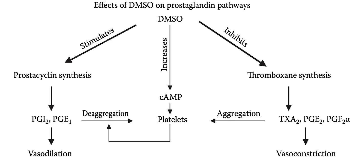
DMSO is often described as a prostaglandin inhibitor (an aspirin-like agent that blocks COX) as for platelets, DMSO selectively inhibits COX-1 and thromboxane A₂ synthase,1,2 cutting TXA₂ production and in cholesterol-fed rabbits, DMSO lowered arterial atherosclerosis, an effect attributed to its reduction in platelet thromboxane B₂ (a stable marker of TXA₂) while prostacyclin was untouched.1 So, since DMSO raises PGE₁ and cAMP while decreasing thromboxane and fibrinogen,1 it shifts the balance towards reducing clotting and dilating vessels.
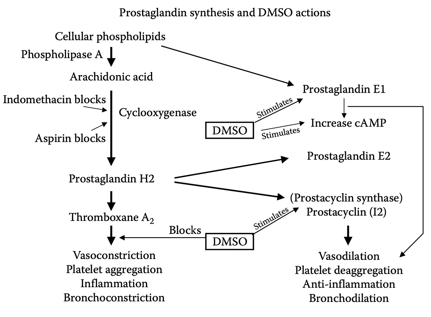
Note: the fuller picture is more complex than “DMSO blocks COX,” as data shows DMSO is not uniformly a prostaglandin-synthesis inhibitor. In one study it increased arachidonic-acid oxidation rather than inhibiting it, didn’t suppress thromboxane, and led its authors to propose it acts as a reducing cofactor rather than a blocker.1 Another found DMSO had no effect on thromboxane yet stimulated PGE₂ and concluded DMSO’s anti-inflammatory action doesn’t run through an arachidonic-acid metabolism at all.1 On skin, DMSO releases prostaglandins and arachidonic acid into the tissue1 (much as histamine does), while in cultured aortic endothelial cells it actually inhibited prostacyclin.1 All of this argues that DMSO doesn’t switch the eicosanoid system off but rather rebalances it, with the net effect in platelets and vessels tilting toward the antithrombotic, vasodilatory side (consistent with DMSO’s broader tendency to normalize dysregulated systems rather than force them one way). Likewise, as we’ve seen before, the effect is also context-dependent, as in some models DMSO adds nothing, and did not improve sevoflurane’s cardioprotection when tested on its own.
Finally, DMSO has also been used to therapeutically deliver beneficial prostaglandins. In one report, topical PGD2 combined with DMSO was used to save digits (fingers or toes) that would otherwise be lost from acute inoperable thrombosis,1 and has been used to deliver agents that act upon this system (e.g., tetrahydroxystilbene⬖1 and curcumin⬖1 to relax the superior mesenteric arteries and arterioles).
Raising cAMP & cGMP
Two closely related molecules cyclic AMP (cAMP) and cyclic GMP (cGMP), once elevated, in platelets, reduce their aggregation and in vascular smooth muscle trigger vessel relaxation and widening.
DMSO raises cAMP through both increasing its production and by slowing its breakdown. On one hand, DMSO directly stimulates adenylate cyclase,1,2,3 the enzyme that synthesizes cAMP; in heart tissue this stimulation was maximal at around 10% DMSO, comparable in magnitude to the beta-agonist isoproterenol and additive with it—yet DMSO’s effect was unaffected by α- or β-adrenergic blockade, meaning it works independently of the adrenergic receptors those drugs act on.1 DMSO also was found to inhibit cyclic AMP phosphodiesterase1 (the enzyme that degrades cAMP), which would further raise cAMP levels.
As such, in platelets DMSO significantly raises both cAMP and cGMP (through a channel entirely separate from its COX-1 inhibition), further supporting its antiplatelet effects.1 In blood vessels, DMSO likewise raises cGMP and relaxes the vessel wall through a nitric-oxide/cGMP-dependent pathway.1
Note: like DMSO’s other effects, this one is concentration-dependent and not universal—at lower concentrations or in certain tissues DMSO showed no measurable effect on the cyclase system.1,2,3
Opening Blood Vessels (Vasodilation)
Blood flow is controlled by smooth muscle surrounding the lining (endothelium) of the blood vessels, which tightens to restrict flow. As such, DMSO’s ability to relax that muscle provides another key way for it to restore tissue circulation. DMSO’s circulatory-promoting properties turn up across nearly every vascular bed studied (e.g., aorta, coronary, pulmonary, mesenteric, renal, cerebral, umbilical, and saphenous) both relaxing vessels by itself and serving as the medium through which other vasodilators are delivered without impeding their effect.1,2,3,4
In the aorta, DMSO produced a concentration-dependent relaxation of contracted aortic rings through an endothelium-dependent pathway (via nitric oxide and cGMP), and an endothelium-independent one that inhibits calcium channels in the smooth muscle and reduces the muscle’s sensitivity to calcium (partly via Rho-kinase).1,2 Likewise, in another study, DMSO relaxed aortic rings by 42-99% and renal arteries by 80% (in part through voltage-gated potassium channels),1 in another DMSO relaxed mesenteric artery preparations, and in drug references, vasodilation is one of dimexide’s (DMSO) recognized pharmacological properties.1
Note: since one of DMSO’s relaxing effects acts directly on smooth muscle, it likely can work on damaged vessels where the endothelium is no longer responsive to stimuli.
Additionally, part of DMSO’s local vasodilation is histamine-mediated—the same mechanism behind the warmth and flushing people often feel where DMSO is applied. Importantly, this is a controlled dilation rather than tissue injury, as when DMSO was injected into a limb, it markedly increased lymph flow while the protein content of that lymph fell (the opposite of what happens with a burn or a caustic agent, where protein rises) and tissue examination showed only vasodilation and occasional edema, but no cell necrosis (death).1 DMSO hence opens vessels and shifts fluid without damaging the tissue.
Note: combined with hydrogen peroxide as an oxygen source, topical DMSO raised skin-flap survival to 92% versus 71% in controls, an effect the authors attributed partly to DMSO’s vasodilatory, histamine-like action.1
DMSO and Nitric Oxide
The classic way a blood vessel dilates is that the endothelium produces nitric oxide, which diffuses into the surrounding smooth muscle and triggers relaxation there through cGMP. For this reason, many integrative cardiovascular health approaches focus on supporting endothelial nitric oxide production.
Much of DMSO’s own vasorelaxation runs through the nitric oxide/cGMP pathway as in isolated aorta, DMSO’s relaxation is blunted by removing the endothelium or by blocking nitric oxide synthase with L-NAME, while conversely, DMSO raises tissue cGMP the downstream marker of nitric oxide signaling1 and at 10% redistributed intracellular nitric oxide into mobile membrane vesicles.1
Furthermore, since DMSO effectively scavenges reactive oxygen species (which cripple the endothelium’s ability to make and use nitric oxide), DMSO is able to preserve the ability of blood vessels to dilate (e.g., protected against cigarette-smoke-induced impairment of nitric oxide production and vasodilation in rabbit aortas and human endothelial cells)1 and, from the opposite direction, DMSO's radical scavenging restored an endothelium-dependent, nitric-oxide–mediated relaxation that iron had blocked in aortic rings.1
Simultaneously, DMSO can counteract nitric oxide when it’s excessive, both by scavenging it directly (DMSO scavenges radicals including nitric oxide), as when IV DMSO in horses modestly raised systolic blood pressure,1 and by throttling its production, as when 2.5–3.5% DMSO (but not 1%) cut endothelial nitric oxide output1—an effect reversed by adding extra arginine (a common supplement used to enhance nitric oxide production).
Collectively, this suggests that DMSO normalizes nitric oxide function, both by preserving it when it would otherwise be impaired, but also by counteracting it if it becomes excessive and harmful (as at low doses, DMSO typically does not affect nitric oxide).
Finally, DMSO has also been used to deliver agents which preserved or restored nitric oxide mediated relaxation such as resveratrol,⬖1,2 hesperidin,⬖ (reducing hyperhomocysteinemia-induced cognitive deficits),1 thymoquinone,⬖1 red-clover isoflavones,⬖1 genistein, ⬖1,2,3,4 (e.g, for fructose-fed hypertension1) methylated quercetin flavonoids and phenylbutanoids,⬖1 Bacopa monnieri flavonoids and saponins,⬖1 tetrahydroxystilbene glucoside,⬖1 astragaloside IV,⬖1 cinnamaldehyde,⬖1 Jasminum sambac extract,⬖1 ursolic acid,⬖1 Acer okamotoanum sap,⬖1 phloridzin,⬖1 adenosine,⬖1 a Periplaneta americana metabolite,⬖1 a polyphenol nutraceutical,⬖1 the soluble guanylate-cyclase activator cinaciguat,1 the PPAR-γ agonist rosiglitazone,1 17β-estradiol,1,2 a STAT3 inhibitor,1 a selective adenosine kinase inhibitor,1 a PPAR-α agonist,1 and an angiotensin peptide analog.1
Counteracting Oxidative Constriction
A large body of literature shows DMSO’s free-radical scavenging (discussed below) protects a wide range of blood vessels from the constriction and endothelial dysfunction caused by oxidative stress, hence keeping vessels from closing down:
In the aorta, it nearly completely prevented ROS-induced vasoconstriction and preserved endothelial and contractile function1 and attenuated hydroxyl-radical- and H₂O₂-induced contractions,1 and it protected platelet-mediated, endothelium-dependent relaxation from hydroxyl-radical damage.1
In coronary arteries, it protected endothelium-dependent relaxation from radical-induced dysfunction,1 and it preserved CGRP-mediated neurogenic relaxation of arteries under oxidative attack.1
In pulmonary and cerebral vessels, it reduced hypoxic pulmonary vasoconstriction by lowering oxygen-radical release,1 and, as a reducing agent, inhibited and reversed the vasoconstriction that oxidizing agents such as peroxide and silver nitrate induce in rat aortas and dog basilar arteries.1,2
Lastly, DMSO also shielded endothelial cells from the injury driven by infection and its toxins, including bacterial endotoxins1 and Pseudomonas aeruginosa1 products.
Note: DMSO’s direct vasorelaxation is concentration-dependent so at the higher concentrations DMSO reaches its own clear relaxing effect, but at the low concentrations used in many solvent experiments, it frequently shows no measurable effect on vascular tone.1,2,3
Free Radical Scavenging
Free radicals are atoms or molecules that contain one or more unpaired electrons, which makes them highly reactive and potentially damaging to tissues such as fats, proteins, and DNA. DMSO’s most well-recognized property is its ability to scavenge (neutralize) free radicals—particularly the hydroxyl radical, one of the most damaging reactive species, of which DMSO is among the best-known scavengers (there are 135,000 results on Google Scholar for “DMSO radical scavenger”). Its reaction with the hydroxyl radical, in fact, is so reliable that DMSO is used as a standard molecular probe for detecting and quantifying hydroxyl radicals in biological systems as the hydroxyl converts DMSO into a stable, easily measured marker.1,2
Note: two of the most commonly observed (and studied) scavenging effects of DMSO are its reduction of free-radical-induced lipid peroxidation and its preservation of the body’s own antioxidant enzymes.1 Additionally, DMSO’s radical scavenging is dose dependent,1 can directly suppress hydroxyl-radical production in solution,1 and delivers vascular protection other approaches cannot—in a cobra-venom model of complement-driven vascular injury, it prevented the microvascular damage (capillary congestion and leukocyte plugging) that cyclooxygenase inhibitors, an iron chelator, and a thromboxane synthetase inhibitor all failed to touch.1
Since the thin lining of the blood vessels (the endothelium) is highly vulnerable to oxidative damage, DMSO’s ability to scavenge radicals has shown a variety of key therapeutic effects including:
•Preventing endothelial dysfunction and preserving vessel dilation.1
•Protecting aortic endothelial cells from radical-triggered programmed death.1
•Blocking the reactive-oxygen signaling that switches on the adhesion molecules (ICAM-1, VCAM-1, E-selectin) endothelial cells use to catch and recruit inflammatory white cells (detailed here) largely by inhibiting the master inflammatory switch NF-κB—thereby preventing those white cells from sticking to the vessel wall, plugging the smallest vessels and causing the microstrokes which underlie many illnesses (e.g., vaccine injuries).
•Blocking the radical-driven stiffening of endothelial cells that would otherwise impair blood flow.1
As such, many studies have shown DMSO protects the endothelium from oxidative stress and free radical injury.1,2,3,4,5,6,7,8,9,10, 11,12,13,14,15,16,17,18,19,20, 21,22,23,24,25,26,27,28,29,30, 31,32,33,34,35,36,37,38,39,40, 41,42,43,44,45,46,47,48,49,50, 51,52,53,54,55,56,57,58,59,60, 61,62,63,64,65,66,67,68,69,70, 71,72,73,74,75,76,77,78,79,80, 81,82,83,84,85,86,87,88,89,90, 91,92,93,94,95,96,97,98
In addition to neutralizing the free radicals that constrict vessels and injure the vessel wall, DMSO protects against a second wave of radical damage: when a vessel’s blood flow is cut off and then restored, the oxygen-starved (ischemic) tissue converts the returning oxygen into a burst of radicals, creating a “reperfusion injury” that often exceeds the damage from the initial blood loss. As such, in virtually every part of the body (particularly the central nervous system), DMSO has been repeatedly shown to protect organs and tissue from oxidative stress and otherwise devastating reperfusion injuries.1,2,3
For example, DMSO:
•Significantly reduced the microcirculatory vascular leakage caused by a radical-generating system in hamster cheek pouches.1
•Reduced vascular permeability and edema in thermally burned skin.1
•Protected dermal microvessels from burn injury.1
•Reduced vascular permeability, edema, and tissue injury in gut and mesenteric ischemia-reperfusion, performing comparably to the antioxidant enzymes superoxide dismutase and catalase.1,2,3,4,5,6,7,8
•Reduced damage in hemorrhagic shock and ischemic gastric injury.1,2,3,4,5,6,7,8,9,10
•Protected the lung and pulmonary vasculature from radical injury.1,2
•Prevented the secondary lung injury that radicals inflict on distant organs after a period of hind-limb ischemia.1
•Reduced endotoxin- and shock-driven organ injury.1
•Improved skin-flap and free-tissue survival by blunting reperfusion radical injury.1,2
•Protected the liver during cold-ischemic preservation.1,2
•Used to counteract the reperfusion radical injury implicated in equine laminitis.1,2,3,4
Note: in many cases, these protective effects are dose-dependent (e.g., in a rat limb ischemia-reperfusion the dose used only gave partial protection,1 while in a lung toxin model, a fairly high dose was needed to provide protection1).
Protecting the Endothelium
The innermost surface of the endothelium is coated with the glycocalyx, a fragile, sugar-rich gel layer that regulates permeability, shields the cells from shear, and keeps clotting and inflammation in check. In shock, sepsis, and reperfusion, this layer is stripped away (”shed”), and its loss is now recognized as an early, pivotal event in vascular collapse.
Note: sulfates are one of the most effective biological molecules for restoring (vital) zeta potential. The glycocalyx is structured so that abundant sulfates on it form a (continually regenerating) liquid crystalline gel around the glycocalyx which repels other substances from entering the endothelium and independently drives blood circulation (all of which is detailed here)—making the glycocalyx one of the most critical components of cardiovascular health.
Many studies, in turn, show DMSO protects the glycocalyx along with the underlying endothelium:
•In a mouse model of LPS-induced acute respiratory distress, systemic DMSO preserved the pulmonary endothelial glycocalyx (staining intensity roughly four times that of untreated injury), cut protein leak into the airspaces, and reduced inflammatory cell counts; in human endothelial cells it prevented glycocalyx shedding by blocking the enzyme (a matrix metalloproteinase) that cleaves it from the cell surface.1
•In a rat hemorrhage-resuscitation model, DMSO reduced glycocalyx shedding and the accompanying coagulopathy.1
•At the level of the underlying matrix, DMSO restored the normal structure of heparan-sulfate proteoglycan in endotoxin-exposed endothelial cells,1 and prevented endotoxin-induced lung protein leak while preserving the endothelium’s ability to relax.1
•DMSO also inhibited apoptosis in nutrient-deprived human endothelial cells by promoting DNA replication and survival,1 and protected them under oxidative stress by raising heme oxygenase-1 and reducing apoptosis through several cytoprotective pathways.1
•In the equine ascending colon, DMSO protected against the capillary permeability changes caused by ischemia-reperfusion injury.1
Note: DMSO has also been used in combination with a variety of agents to promote endothelial growth and repair (e.g., in studies of endothelial proliferation, migration, tube formation, differentiation, and progenitor-cell-driven revascularization). Agents combined with DMSO to create these effects on the endothelium include: curcumin, ⬖1 resveratrol,⬖1 luteolin,⬖1 kaempferol,⬖1 diosgenin,⬖1 icariin,⬖1 arnebin-1,⬖1 celastrol,⬖1 dihydroartemisinin,⬖1 alkannin,⬖1 sulforaphane,⬖1 ginsenoside Rg3,⬖1 Illicium henryi extract,⬖1 dracorhodin,⬖1 onychin,⬖1 Echinacea extract,⬖1 13-cis retinoic acid,1 fluvastatin,1 simvastatin,1 atorvastatin,1 dihydrotestosterone,1 recombinant proteoglycan-4,1 sirolimus1 rapamycin,1 a demethylating agent,1 a Kir2.1 blocker,1 or a JAK2 inhibitor.1
Current Stroke Management
Roughly 3.1% of adult Americans have experienced a stroke (a figure I expect to rise from the COVID-19 vaccines). Each year, this translates to about 800,000 people in the United States having a stroke, and in 2022, 165,393 died (making it the fifth most frequent cause of death in the United States), with between 20-40% of survivors experiencing long term disability from the stroke.
Because of the harm strokes pose to society, and the rate at which brain tissue deteriorates once its blood supply is lost, the medical system emphasizes doing everything that can be done to treat strokes as soon as possible. Unfortunately, there is a fundamental limitation to how strokes are addressed which makes it impossible to ever eliminate the immense toll strokes place upon society.
This is because three types of strokes exist, ischemic strokes (which are typically caused by a blood clot obstructing the artery), a brain hemorrhage (where bleeding is both directly toxic to sensitive brain tissue and creates large blood pockets that compress and damage the brain), and transient ischemic attacks (TIAs)—which are not classically considered strokes.
Note: TIAs occur when someone shows clinical signs of a stroke that resolves on its own (and typically does not have stroke imaging findings). I believe TIAs often represent microstrokes occurring (as the microclots are too small to be seen with conventional brain imaging and typically resolve on their own)—a subject I went into much more detail here as these visually detectable microstrokes frequently underlies vaccine injuries and many other chronic illnesses.
The essential challenge with managing strokes is that it’s nearly impossible to reliably differentiate an ischemic stroke from a hemorrhagic stroke without imaging (we’ve tried for a longtime, and while there are a few diagnostic signs that suggest one or the other, they are not reliable enough for the degree of certainty required). This matters because opposite approaches are taken to ischemic strokes and hemorrhagic strokes—with ischemic strokes a powerful clot busting medicine (tPA) is used to restore circulation, whereas with hemorrhagic strokes (13% of strokes in the developed world), a variety of steps are taken to reduce further bleeding in the brain (which can include brain surgery).
As such, when a stroke occurs, while it is urgent to treat it as soon as possible (as “time is brain”), the standard approach (tPA) cannot be used until a CT scan has ruled out a hemorrhagic stroke (as giving tPA for a hemorrhagic stroke is devastating or lethal). Because of this, definitive stroke treatment cannot begin until it has been recognized, an ambulance gets them to an ER, a CT scan has been completed, a diagnosis is made, and that diagnosis reaches the treating physician—all of which, despite the best efforts by the medical system to expedite the process, often takes hours.
Worse still, the statistics on tPA (approved in 1996 and still the only FDA approved treatment for ischemic strokes) aren’t actually that good. Presently, tPA is only approved to be given within 3 hours of a stroke starting (as its likelihood of benefitting a patient decreases with time), and in practice, it is often given up to 4.5 hours after symptoms start (since some degree of benefit still exists).
When that window is met (which only happens about 25% of the time and ultimately results in roughly 1.8%-8.5% of ischemic stroke patients receiving tPA), the existing data shows that only 13% percent of patients who receive tPA significantly benefit from it (39% return to normal, compared to 26% who would return to normal without treatment), with an additional 19% of tPA users experiencing some degree of improvement (but not a full recovery) from it.
Worse still, tPA can cause significant bleeding, which is sometimes minor (e.g., gum bleeding), but also carries a 6.4% risk of a symptomatic brain bleed, and a 1.6% risk of a serious systemic hemorrhage (along with other issues such as a 1.3% to 5.1% risk of angioedema and tPA frequently causing reperfusion injuries). In turn, many risk factors exist for the increased bleeding (e.g., a few common risk factors can lead to a 33% chance of tPA causing a fatal bleed), and there have been many lawsuits for either giving or not giving tPA to a stroke patient. Additionally, tPA is a poor choice for larger obstructions (e.g., one within the internal carotid artery), which instead must be physically removed. In short—many ICU doctors I know are quite hesitant to use tPA as they have seen cases where it dramatically improved patients, many where it did not do anything, and quite a few disasters (especially in the early days of the therapy where it was used for heart attacks and then often caused the patient to have a fatal or debilitating brain bleed).
Note: the best data exists for tPA being injected directly into the obstructed artery with interventional radiology. Unfortunately, while many premier institutions offer this, it is a specialized procedure that is not available at most hospitals.
Finally, there is essentially no therapy for recovery from stroke—which in short explains why stroke is the second leading cause of death and the third leading cause of disability worldwide.
DMSO and Stroke Management
As the previous section shows, there are insurmountable limitations to how strokes can be managed. DMSO, however, completely changes this equation as:
•It effectively treats ischemic strokes.
•It partially treats hemorrhagic strokes.
•It carries no known risk of worsening a hemorrhagic stroke.
•It can be easily administered at home or on an ambulance.
•In addition to addressing the circulatory issues, independently protects brain tissue from stroke damage.
•Prevents the reperfusion injuries which follow strokes.
•Can heal damaged brain tissue after a stroke.
As such, DMSO makes it possible to eliminate the multi-hour delay typically required for stroke treatment, often is a safer can be a more effective stroke treatment option than the conventional alternatives, and provides one of the only existing options to heal the permanent disability that follows a stroke.
This is why researchers like Jack de la Torre MD devoted their entire careers to producing a robust body of evidence DMSO could treat strokes, as they knew it was unlikely any other agent would ever be able to solve the ischemic-hemorrhagic stroke dilemma.
Note: many readers have reported to me successfully treating strokes at home or on the way to the ER with DMSO and hence, due to increasing skepticism towards hospital care,1 have advocated for home DMSO in lieu of going to the hospital. This is extremely unwise, as if a significant hemorrhagic stroke is occurring, DMSO cannot work as the primary treatment. Rather, the correct course is to use DMSO on the way to the hospital, and then if the whole thing is written off as a TIA (which many people have shared happened to them after they used DMSO, it resolved the stroke midway to the ER, and ER could then not decide if a stroke had happened), simply be grateful you had the optimal outcome when things could have easily been much worse. In short, the existing management we have for strokes is correct, and DMSO should be viewed as way to improve the results it yields rather than a replacement for it.
Ischemic Stroke Data
Many stories like these two Archie Scott reported1 exist throughout the DMSO literature:
A Los Angeles school teacher had a major stroke shortly after the start of the Christmas break. She was unconscious on her living room floor. DMSO treatment was started immediately after the stroke. The DMSO was first applied topically to her head within minutes of the stroke. Less than one hour after the stroke she was given DMSO by intramuscular injection. This patient was never taken to the hospital for this stroke. A prominent surgeon who was a family friend told the husband of this patient that it was important to keep her out of the hospital. The surgeon said that even though the treatment was completely legal, it would be difficult to get approval to give the DMSO especially by injection at his hospital.
This patient made a dramatic recovery. She regained consciousness later in the day in which she had her stroke. Treatment continued for the next week. Each day she received two topical applications of DMSO, one intramuscular injection of DMSO, and two doses of one teaspoonful of DMSO in juice. Her condition improved each day. When school resumed after the first of January, this teacher was back in the school teaching the students as if nothing had happened during the Christmas vacation. She continued teaching until she retired, healthy and with no disability.
A lady was in a coma in a convalescent hospital and had been in the coma since her stroke three months ago. She was given little chance of recovery and was expected to remain in a vegetative state until her death.
When I first observed this lady, there was no response to any type of stimulus. She was alive, but appeared lifeless. It was decided that her treatment should be topical DMSO daily….One month after the start of treatment, there were positive signs in the lady. Her brain was starting to respond to the DMSO. The treatment continued, and four months after treatment started this lady was able to return to her home [where treatment continued].
Three years after the start of DMSO treatment this writer returned to visit this patient. At this time the lady was living a normal life, not the life of a stroke victim. She was able to look after the house and walked normally.
The only lingering effect of the stroke was a slight speech defect. At this time she said that her memory was better than that of her husband who had not had a stroke and who was considered to be completely normal.
Note: there are also many reported cases of individuals who took DMSO for musculoskeletal or pain disorders (by far the most common use of DMSO) who then experienced a permanent improvement of stroke symptoms.
Extensive data, in turn, corroborates these remarkable stories.
Human Evidence
Due to the ethical issues with creating and then treating strokes in humans, most of the research in this field has been conducted in animals (which I hold many strong ethical objections to). As such, the only human study has been conducted in ischemic strokes was a 2002 trial, where IV DMSO given with fructose diphosphate twice daily to 11 patients (mean age 65) with acute or subacute ischemic stroke was well tolerated and, when started within 12 hours of stroke onset, left 63% “improved” or “markedly improved” versus 20% of those on standard care at three months, and the benefit held even when treatment began well after the stroke.
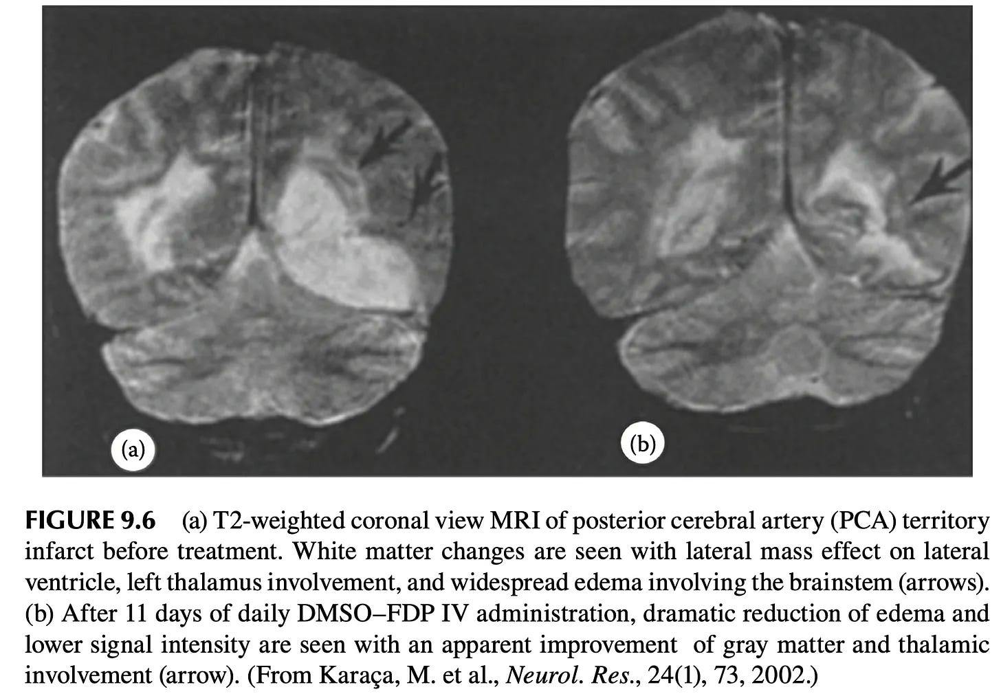
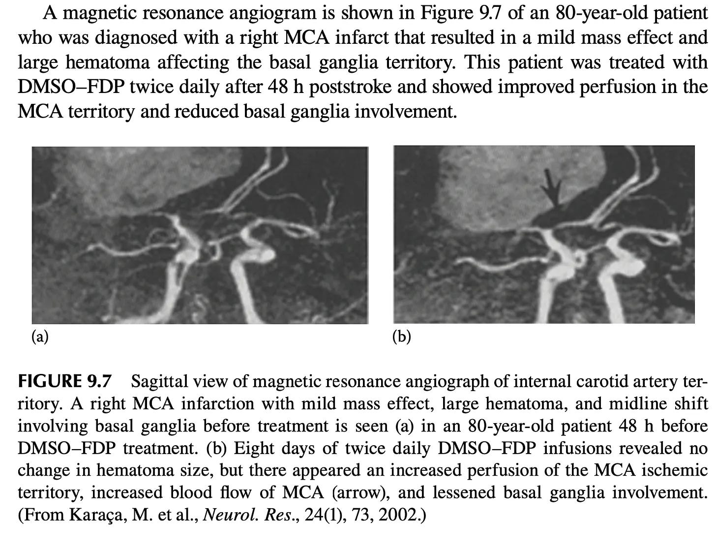
Note: animal studies further corroborate DMSO-FDP value. In rabbits whose brains were driven to flatlined (isoelectric) EEGs by combined hypoxemia, hypotension, and carotid occlusion, DMSO-FDP given after five minutes of electrical silence restored brain activity far faster and let all animals survive with minimal damage, versus only 22% survival (severely disabled) on saline, and in mice given head impacts (another area DMSO helps), DMSO-FDP was the most protective and DMSO alone the second, while FDP and the rest gave no benefit, confirming DMSO was therapeutic. Finally, when DMSO-FDP was given to rats with poor chronic blood flow to the brain (due to carotid obstruction), there was a 54% improvement in visuo-spatial memory1,2 (which is corroborated by dementia patients who’ve improved from DMSO).
•A Russian patent treated perinatal hypoxic encephalopathy in newborns by daily electrophoresis of vitamin E⬖ in DMSO. In three term infants 6–7 sessions produced disappearance of limb tremor (chin tremor remaining only with crying), normalization of muscle tone, return of tendon and support/automatic-walking reflexes, loss of the spontaneous Moro reflex, and restoration of the Babkin reflex, with faster clinical recovery than intramuscular vitamin E⬖ plus standard care.
•One physician reported knowing of a patient of Stanley Jacob’s whose stroke was successfully treated with DMSO.1
Note: I have corroborated through multiple sources that Stanley Jacob successfully treated numerous strokes with DMSO, but I have not been able quantify how many in total were treated or what the success rate was.
Additionally, numerous reviews support DMSO’s use in humans with strokes. These include:
•A 2009 pharmacology review by Jack de la Torre and Stanley Jacob of DMSO in cardiac and CNS damage (arguably the most detailed one in print) showed intravenous DMSO elevated cerebral blood flow, limited infarct volume after middle-cerebral-artery occlusion, preserved the vulnerable CA1 and dentate neurons and facilitated recovery from acute or chronic cerebral ischemia.1
Note: de la Torre was publishing papers on DMSO treating strokes fifty years ago1 and likewise, a 1982 paper1 acknowledged DMSO was being used to treat strokes.
•A 1992 Chinese review that concluded DMSO protects ischemic brain tissue and blocks the secondary pathological processes that follow cerebral ischemia, outperforming mannitol, dexamethasone, and barbiturates as a neuroprotectant.1
•A 2010 Chinese review of DMSO’s effects on the heart and central nervous system (which noted authors had identified over 10,000 articles on the biological effects of DMSO).1
•A 2012 Chinese review classified DMSO as a classic antioxidant with therapeutic effects on ischemia reperfusion injury across many organs (and noted it had already been used clinically abroad for cerebral reperfusion injury).1
•In a 2018 Ukrainian physical-rehabilitation review where DMSO appears as an iontophoresis solvent in post-stroke and disability rehabilitation protocols.1
•A 2022 Russian review from Osh State University argued that drug electrophoresis (driving charged medication ions through the skin with direct current) is among the most valuable physiotherapy methods for cerebrovascular disease (stroke and TIA) and peripheral-nerve disorders. Within it, DMSO is used as a carrier that lets otherwise water-insoluble drugs dissociate and be pushed through the skin. In acute stroke in the early recovery period, this was applied to the eyelids and back of the neck with heparin added for ischemic stroke and iodine substituted for hemorrhagic stroke.1
DMSO Alone in Stroke Models
DMSO has repeatedly been shown to reduce the core injury of an ischemic stroke:
•A rhesus monkey study blocked the MCA for 4 hours, gave DMSO, dexamethasone, or nothing, and then opened the MCA after it had been blocked for 17 hours. DMSO gave significant protection from the severe neurological deficits and loss of arterial blood flow the other two groups developed.

•A squirrel monkey study blocked the left MCA for 4 hours, after which the animals were given a variety of different treatments (e.g., saline, hemodilution, or hyperbaric oxygen at 2 atmospheres). Seven days after treatment, 8 of 10 DMSO treated monkeys were alive (with 2 having mild contralateral muscle weakness), while 75% of those receiving hyperbaric oxygen survived, and just 34% of those receiving hemodilution survived (with the last two groups also having more significant neurological deficits). Finally, combining either of these treatments with DMSO produced slightly worse results than just DMSO alone.
•In experimental canine middle cerebral artery embolectomy, DMSO (or low-dose methylprednisolone) extended the grace period to 6 hours following embolization and protected the cerebral tissue from the injury of ischemia and post-ischemic reperfusion (with no infarcted tissue in DMSO treated animals and a 1.45 cm3 area of infarction in untreated ones).1,2,3,4
•In a canine model of severe cerebral ischemia (where blood flow was cut by 90% for an hour), DMSO given alone as pretreatment allowed EEG activity to re-emerge after recirculation where untreated animals showed none. In a second canine study, in brains subjected to 14 minutes of complete ischemia and then reperfused for an hour, DMSO added to the reperfusion blood lowered a marker of lipid peroxidation (TBAR) back to control levels, restored roughly three quarters of the lost energy stores (ATP and creatine phosphate), cut lactate accumulation by 60%, and produced a marked return of EEG and auditory evoked potentials that untreated brains entirely lacked. Adding a platelet-activating factor antagonist on top of the DMSO further improved mitochondrial energy markers, though the reduction in lipid peroxidation was attributable to DMSO alone.
Note: the first study also found vitamin E⬖ restored brain function, mirroring forgotten Russian research on how to counteract the biological consequences of chronic stress on the brain with DMSO and vitamin E.⬖
•In a canine model of pressure-induced focal ischemia worsened by ethanol exposure (done to model head trauma from a drunk driving car accident), DMSO significantly reduced brain lesion volumes under both normotensive and hypotensive conditions and scavenged the hydroxyl radicals generated from ethanol metabolism.
•When seven different therapies were compared head-to-head in a canine model of severe pressure-induced focal ischemia, intravenous DMSO produced the best neurobehavioral recovery scores and the smallest lesion volumes, with reduced edema, necrosis, and cavitation.
•In a rat transient focal ischemia model done with 90-minute middle cerebral artery occlusion followed by reperfusion (as the MCA is one of the most commonly sites of consequential strokes and hence often blocked to simulate strokes), DMSO given 30 minutes before ischemia dose-dependently reduced cortical and striatal infarct volumes (how much brain tissue was damaged), and at the higher dose also significantly improved neurological motor function (while a low dose produced no benefit).
•In rats, DMSO given 30 minutes prior to MCA occlusion significantly reduced the amount of permanently damaged brain tissue1,2,3 In another, DMSO immediately after occluded MCA blood flow was restored reduced rat infarct size and blood-brain-barrier damage (as measured by MRI), with enhanced protection and reduced MMP-2/MMP-9 activity (enzymes that breakdown the blood–brain barrier) when combined with DPI. Additionally, oral DMSO with vitamins C⬖ and E⬖, given 12 hours after rat MCA occlusion, significantly reduced oxidative stress.
•In rats, DMSO one hour before or after MCA and carotid occlusion significantly reduced rat brain edema and infarct volume. In rats with focal cerebral ischemia DMSO reduced brain water content and lipid peroxidation, preserved Na⁺-K⁺-ATPase and superoxide dismutase activity relative to saline controls, and markedly improved brain edema and ischemic tissue damage.
•In rats subjected to four vessel occlusion (which cuts off almost all blood flow to the brain) DMSO partially reduced mitochondrial malondialdehyde and free fatty-acid accumulation after reperfusion.
•In neonatal (7 day old) rats with hypoxia-ischemia brain damage, DMSO injected into the brain reduced infarct volume and brain injury (particularly within the cortex) along with inhibiting the breakdown of MAP2 and fodrin, suggesting neuroprotection via calpain inhibition.
•In gerbils subjected to carotid ischemia-reperfusion, DMSO, significantly reduced delayed neuronal death and lowered markers of hydroxyl radical activity that accumulates after reperfusion.1 In another study, intraperitoneal 10% DMSO before ischemia cut gerbil mortality from 60% to as low as 14%, reduced the proportion of ischemic hippocampal neurons (from 71% to 46% at the higher dose), and blunted neurological signs.1,2
•In a thrombotic (clot) stroke mouse model, daily DMSO (given for five days after the infarction) prevented the shrinkage and nuclear condensation of injured neurons and, most strikingly, sharply reduced the excessive astrocytic scarring response (gliosis) in the damaged cortex and hippocampus, restoring the astrocytes toward their normal morphology.1,2
•In decapitated mice (with blood flow hence being cut off to the brain), DMSO prolonged the time the animal continued to gasp for breath and produced a dose-dependent relaxation of pre-contracted vascular smooth muscle, pointing to a vasodilatory contribution to its anti-ischemic effect.1 An earlier report found DMSO was protective experimental hypoxic respiratory depression.1
•In a rat model of deep hypothermic circulatory arrest, the DMSO solvent-control group “unexpectedly” showed some of the same organ-protective effects as the active drug (ebselen) it was carrying.
Note: many of these results argue that giving IV DMSO beforehand could reduce the complications of many challenging surgeries (e.g., a coronary bypass). Unfortunately, much in the same way ultraviolet blood irradiation dramatically reduces bad surgical outcomes, neither has been adopted for this purpose.
Beyond reducing the core infarct, DMSO also protects the penumbra. Frequently in strokes, an area will form where blood has been impaired, but brain tissue has not yet died (known as the penumbra and the key target of most stroke management). In a rat stroke study where DMSO was administered an hour after brain blood flow had been permanently cut off, MRI imaging showed that DMSO stopped the region of dying brain tissue from continuing to expand, hence allowing a penumbra (rather than additional dead tissue) to form around the stroke site (particularly within the cortex).
Note: beyond the classic penumbra, groups of cells can also enter a shocked state where their normal functions cease (and they eventually die). As discussed here, this “penumbra” also responds to DMSO (which is one reason tissue often comes back to life following DMSO treatment and why the sooner DMSO is used after a brain or spinal injury, the better the results typically are).
•In guinea-pig hippocampal slices, 0.4% DMSO alone more than doubled the latency to irreversible ischemic depolarization (from 2.9 to 7.1 minutes) under combined oxygen-glucose deprivation while also reducing its amplitude, an effect further augmented when DMSO was combined with nimodipine.
•In rat hippocampal slices, 10 mM DMSO completely prevented the hypoxia-induced drop in serotonin (5-HT) release and directly increased spontaneous release under normoxia (indicating a direct stimulatory action on serotonergic neurons).
Finally, DMSO also potentiated hypoxic preconditioning on its own: given intraperitoneally or directly into the hippocampus, it increased rats’ resistance to severe hypoxia by roughly 79% and 111% respectively over controls (via α7-nicotinic receptors).
Note: a veterinary text1 (along many with other parts of the veterinary literature1) also highlighted DMSO’s edema-reducing, diuretic, and anti-inflammatory effects in cerebral ischemia. Likewise, in veterinary neurology, DMSO was used therapeutically or as a carrier in temporohyoid osteoarthropathy with acute neurological signs1, post-anesthetic air embolism with neurologic sequelae in a horse1, critically ill neonatal foals1, a filly’s watershed cerebral infarction1, and a 2-day-old filly with hypoxic-ischemic encephalopathy given IV DMSO.1
How DMSO Protects the Brain
In addition to the extensive research showing that DMSO restores blood circulation and prevents reperfusion injuries in those (previously ischemic) tissues, numerous studies have shown DMSO mechanistically protects the brain from strokes:
•In anesthetized cats, DMSO significantly enhanced brain oxygenation (particularly in the caudate nucleus).
•DMSO was shown to preserve the neurological function of hippocampal brain tissue samples once their oxygen or glucose were withdrawn (with similar results seen in this study).
•When infused directly into the rat brain, DMSO alone reduced ischemia-induced (blood loss induced) extracellular glutamate (the excitotoxin that drives much of the neuronal death in a stroke) by 51% and modestly preserved the vulnerable hippocampal CA1 neurons relative to controls.
•DMSO down-regulated aquaporin-4 expression (a key driver of the brain swelling that follows an occlusion) after MCA occlusion in the rat.
•DMSO was found to protect the integrity of the blood-brain barrier integrity after MCA occlusion.
•Diazoxide and DMSO prevented the learning dysfunction and brain damage that follow carotid artery occlusion (with DMSO alone restoring) Morris-water-maze learning and preventing COX-2-positive neuron loss in the dentate gyrus.
Reader Stroke Reports
Corroborating DMSO’s benefits for strokes, numerous readers here shared the following with me:
•A medicinal chemist with a background in neuroscience, recognized his own stroke the moment it began: “Right after waking up I went to the toilet and noticed that I had lost control of my palm. I walked to bed and I froze, unable to lay down.” He had his wife get him diluted DMSO, drank most of it over the next hour, and went to the hospital—by which point he had lost speech (Polish entirely, English partially), leaving him, a Pole, to communicate in English in a Polish hospital. “I credit DMSO for not losing movement ability,” he wrote. His speech recovered well enough that he returned to full-time work as a scientist six weeks later, and he now drinks a small maintenance dose daily.1
•One week after her husband’s stroke, which had left him with severe pain, vertigo, exhaustion, and partial paralysis of the left side of his face, arm, and leg, a reader discovered this series. She then applied undiluted DMSO to his face, neck, and shoulder two to three times a day; the pain, she reported, “went away almost instantly every time.” Within about two weeks he began walking short distances without a cane. “Only 3 months after the stroke he walked with me unassisted to a friend’s house carrying a guitar, and was able to play the guitar and sing flawlessly while standing with one foot on a chair. My jaw was on the floor... ‘You’re all witnessing a freakin’ miracle right now. This man had a stroke three months ago, and now he’s playing his guitar like nothing happened!’”1
Thanks for saving my life. I had a stroke recently, and because of your article on dmso and strokes, was given some by my partner. Three minutes later I was OK again. I continue to read your articles daily.1
I have been in health chats where twice now, folks were in the chat and were having a stroke, they both had DMSO on hand & took 1 oz it both were stopped within 10-15 min and reversed any damage.1
I just recently had a stroke but used DMSO immediately ( I had my wife apply it in the hospital within hours of my stroke) and I’m about 95% back to normal and hoping to be 100% soon.1
•A wife whose husband had atrial fibrillation and two microclot strokes in 2024 treated a recurrence herself the following year: when both his legs went numb with pins and needles, she applied DMSO and “Within two hours he was 70% better,” and after a second identical treatment “everything was cleared up and did not return” (months later she used the same protocol over his chest for a suspected pulmonary embolism, with the same result).
•Another wife, reported DMSO “Made a world of difference” for her husband’s entire-left-side deficit,1 another reported their husband’s two mini-strokes cleared within a few hours,1 another reported that when her husband woke up unable to speak and with numbness in his cheeks, she began DMSO, and there was “a huge improvement from a week ago” as he was speaking in complete sentences most times.1
•A reader who saved her mother from a third stroke shared “It was miraculous!”1; another reported it saving a family member from a stroke.1 Likewise, a reader whose father began showing stroke symptoms wrote: “Thankfully I had some 99.9% DMSO on hand. The improvement was remarkable….“The Lord might have just used you to save my dad’s life.”1
Interestingly, many readers have reported stories matching my own experience of giving someone DMSO and then having the ER be confused because they could not find any signs of stroke:
•After a reader woke at 4:30 a.m. and found his left side would not respond when he tried to walk stated he immediately knew to use his DMSO and over the next 4 hours “My walking returned to normal but my reach and grab with my left arm/hand would not allow me to pick green beans in my garden.” He hence called 911, walked down his own hill to greet the firemen, and talked the EMTs out of taking him; the next morning his clinic’s nurses sent him to the hospital by ambulance, where monitoring, a CT scan, and echocardiograms found “no evidence of stroke,” and his remaining left-hand impairment recovered over two weeks with self-directed therapy. His conclusion: “In every interaction with medical personnel I strongly recommended that DMSO should be the first/immediate action when stroke symptoms are detected. Waiting for an ambulance or an emergency room visit to respond is a tragic waste of time.”1
•A 76-year-old man in excellent health had a nearly identical experience 36 hours after giving a double-red blood donation: he woke at 4 a.m. with trouble walking and gripping on his left side, and applied DMSO based on what he had read here. Hospital testing “has not found/confirmed stroke,” and he reported rehabbing himself until symptoms had “disappeared 97%.”1
•Another reader who was already familiar with DMSO and kept it on hand took it during an apparent stroke: “The ER doc came in to tell me that I had not stroked, despite speech impairment and other signs. I told him I took DMSO.”1
•The reports extend well beyond the acute window into recovery from established strokes. One reader’s husband, who had suffered eight strokes, takes DMSO three times a week; two years after his last stroke, in her words, “recovery great, he has some aphasia but still thinking great speaking to others although he’s an introvert and has lost some confidence in himself he is still living life to its fullest playing with grandkids gardening home renovations and going to the gym and doing a full work out which includes 30 chin ups!”1 A caregiver eight weeks into treating a stroke patient (who noted the changes “began almost immediately, after the very first treatment”) observed a cascade of gains: stronger voice and markedly improved speech, dramatically improved swallowing (he could again eat normal-sized meals, and even swallow while the TV was on, previously a choking hazard), self-feeding with the affected hand; improved energy, strength, and balance; and, “the most recent and undeniable change,” the return of his sense of taste. On one lucid day he zipped up his own jacket—notable, she wrote, “because the fine motor skills in his right hand were quite impaired by the stroke” and “even he commented at the time that it was remarkable.”1 Another reader watching his mother’s recovery described her damaged right side visibly “waking up”, regaining feeling and strength until one day she exclaimed, “Hey look, I’m sanding with my right hand!”1
•Others report gains across the full range of stroke severity. One reader had a large stroke that “affected every joint on my right side” and used DMSO with CBD: “One year later? Only my knee and two fingers (pinky and ring) are still ‘lazy’ but each month I notice an improvement... I couldn’t write but I can now. I’ve learned to type with 8 fingers but the other two are still improving.”1
•Another reader began giving her 72-year-old husband DMSO six weeks after his stroke; over two and a half weeks “he improved and slowly regained more function so that he could swim again,” and a later course brought further gains. She noted the oral route seemed to act faster, though its garlic-like odor kept him homebound—so he switched to applying DMSO nightly to his temples, forehead, and neck, on which he continued to slowly improve.1
•A 65-year-old who’d had a series of small strokes reported that after starting DMSO the improvement in mental acuity was “amazing” alongside whole-body pain relief;1 a physician who ran an IV chelation and ozone clinic recalled an “amazing recovery” in a stroke patient given IV DMSO;1 and caregivers successfully treating loved ones after strokes (e.g. “a very bad stroke1”) or an ischemic stroke and reporting steady, believable progress.1,2,3
•A grandfather who nebulized DMSO for decades survived multiple strokes and “amazed doctors by how quickly he recovered,” living to 94.1 Another reader stated flatly that DMSO saved him from a stroke a decade ago.1
Finally, many readers describe using oral DMSO as ongoing stroke prevention. One was having strokes two or three times a week until he started DMSO (originally bought for sciatic nerve pain): “the strokes stopped happening... once it is in the blood stream it works everywhere.”1 Another also reported DMSO stopped their strokes,1 Likewise, a diabetic who’d had two strokes from clotting that monitoring his own blood under a microscope, found daily oral DMSO kept his red cells out of rouleaux formation and “the clots at bay.”1
Note: these readers were almost certainly suffering from what would be diagnosed as “TIAs” and the fact that both DMSO prevented their recurrence and that one reader could link this to DMSO eliminating blood sludging argues for my theory many TIAs are actually microstrokes triggered by things like zeta potential disruption.
In short, I hope all of this (and what is to come) makes it clear why I feel so strongly about DMSO becoming a standard therapy for strokes. Nonetheless, as discussed earlier, it is absolutely critical you do not avoid emergency care just because DMSO is available and rather treat it as complementary therapy as you wait for conventional care.
DMSO Stroke Combination Therapies
Since DMSO is both a non-toxic solvent and able to deliver a variety of agents (e.g., through the skin), that rare combination results in it being used as a “vehicle control” in many studies. In these studies, DMSO is typically assumed to be inert (and hence not independently tested for an effect), but in some cases is tested, where it often yields a therapeutic effect (which I’ve noticed authors often do not disclose). As a wide number of agents combined with DMSO all yield therapeutic effects similar to those seen with DMSO, this suggests that DMSO plays a direct role in the effects observed, particularly since DMSO frequently potentiates the agents it is combined with. This dynamic I believe likely accounts for why many agents that succeed in preclinical studies (where DMSO is also used) fail in clinical studies where DMSO is not also used.
Note: replication failure is one of the largest problems in science.
As such, I include these combinations here (most of which were used for focal brain ischemia) both to highlight the shared effects seen across many agents (that potentially are due to DMSO) and to provide insights to readers looking for additional treatment strategies. To make these section easier to skim, all natural agents are marked with a ⬖.
Natural agents
Polyphenols & flavonoids — curcumin⬖ (reduced infarct size and cell death)1,2,3,4,5,6,7,8,9,10,11,12,13,14; resveratrol⬖ (reduced infarct size and preserved neurons, including in elderly and recurrent-stroke rats)1,2,3,4,5,6,7,8,9,10,11; baicalein⬖ (reduced infarct size and cell death)1,2; chrysophanol⬖ (reduced infarct size and cell death as a pre-treatment)1,2; quercetin⬖ (reduced infarct size)1,2; chrysin⬖ (improved movement and memory and reduced oxidative stress)1,2; nordihydroguaiaretic acid⬖ (reduced neurological deficits and infarct size)1; apigenin⬖ (reduced inflammation)1; mangiferin⬖ (reduced neurological deficits when given before ischemia)1; shikonin⬖ (reduced infarct size and oxidative stress)1; trifluoro-icaritin (reduced neurological deficits and infarct size)1.
Alkaloids & related plant compounds — berberine⬖ (reduced infarct size and cell death)1,2,3,4; evodiamine⬖ (reduced infarct size)1,2,3; rutaecarpine⬖ (improved movement and raised neurotrophic factors)1,2,3; cynandione A⬖1; vinpocetine⬖ (reduced infarct size and inflammation)1; sinomenine⬖ (reduced inflammation)1; piperine⬖ (reduced injury)1; carvacrol⬖ (reduced injury)1.
Terpenoids, saponins & quinones — astragaloside IV⬖ (protected neurons and reduced cell death)1,2,3,4,5; celastrol⬖ (reduced neurological deficits and infarct size)1,2; the cembranoid 4R⬖ (reduced infarct size and cell death)1; ginsenoside Rb1⬖ (reduced neuronal death)1; bilobalide⬖ (reduced neurological deficits and infarct size)1; tanshinone IIA⬖ (reduced infarct size when given after stroke)1; sulforaphane⬖ (reduced infarct size and inflammation)1; triptolide⬖ (reduced infarct size, swelling, and neurological deficits)1; picroside⬖ (protected mitochondria and reduced cell death)1; ursolic acid⬖ (reduced infarct size and improved neurological scores)1; Z-guggulsterone⬖ with boswellic acid⬖ (improved neurological function and promoted new blood-vessel growth)1; salvinorin A⬖ (reduced inflammation and swelling and protected the blood-brain barrier)1,2,3,4; herkinorin (reduced brain swelling and improved neurological function)1,2.
Whole-herb & multi-herb extracts — kava extract⬖ (reduced cell injury and inflammation)1; Gastrodia elata extract⬖ (reduced infarction by shifting immune cells toward healing)1; Ptychopetalum olacoides extract⬖ (protected brain tissue from oxygen deprivation)1; Angelica sinensis extract⬖ (improved neuronal survival and regrowth)1; Ginkgo biloba extract⬖ (prolonged survival and reduced injury)1,2; Viola spathulata extract⬖ (reduced infarct size)1; Zhenbao Pill⬖ (reduced inflammation and cell death)1; Qing-Nao-Tong-Luo Recipe⬖ (reduced infarct size and improved neurological function)1; Tongxinluo⬖ (protected the blood-brain barrier and circulation)1; Shenmai injection⬖ (reduced deficits and infarct size)1.
Other natural compounds — stilbazulenyl nitrone⬖ (was highly neuroprotective as an antioxidant)1; docosahexaenoic acid⬖ (protected brain cells from oxygen deprivation)1.
Synthetic & pharmacological agents
Kinase & MAPK-pathway inhibitors — an ERK inhibitor (improved survival and protected mitochondria after cardiac arrest, and reduced infarct size)1,2,3,4,5,6; a PI3K inhibitor (reduced injury in diabetic brain injury)1; a p38 MAPK inhibitor (reduced cell death and swelling and improved neurological function)1,2,3,4,5,6,7,8; a JNK inhibitor (protected neurons and improved survival)1,2,3; rapamycin (reduced infarct size)1,2,3,4; Mdivi-1 (reduced oxidative stress and cell death)1,2; a STAT3 inhibitor (reduced injury)1; anisomycin (improved neuronal survival)1; a PTEN inhibitor (promoted neuronal repair)1; HIF prolyl-hydroxylase inhibitors (induced protective autophagy)1; bosutinib (reduced infarct size and inflammation)1; a TAK1 inhibitor (improved neurological scores and reduced infarct size)1; 5’-deoxy-5-iodotubercidin (reduced infarct size by up to 57%)1; 2-Cl-MGV-1 (promoted new blood-vessel growth)1.
Cell-death & protease inhibitors — caspase inhibitors (reduced infarct size and cell death)1,2,3,4,5; cathepsin inhibitors (reduced infarct size and cell death)1,2,3,4; a Notch/γ-secretase inhibitor (drove stem cells toward neurons and aided post-stroke regrowth)1,2; necrostatin-1 (reduced necroptosis and improved myelination and memory)1,2,3; cyclosporin A (protected neurons)1; a calpain inhibitor (reduced cell death and injury)1,2; a PARP inhibitor (reduced infarct size)1; thalidomide (reduced infarct size and cell death)1.
HDAC & epigenetic inhibitors — an HDAC inhibitor (trichostatin A) (reduced infarct size and inflammation)1,2,3; an HDAC inhibitor (SAHA) (reduced infarct size by up to 57%)1,2; an HDAC inhibitor (MS-275) (reduced infarct size)1; an HDAC inhibitor (TSA) (reduced infarct size and inflammation)1; an EZH2 inhibitor (improved coordination and reduced injury)1.
Nuclear-receptor & metabolic modulators — pioglitazone (reduced neurological deficit, swelling, and inflammation)1,2,3,4; all-trans retinoic acid (reduced infarct size as a post-treatment)1,2,3; a PPARγ inhibitor1,2; LXR agonists (reduced infarct size)1; rosiglitazone (reduced infarct size, and in DMSO cut it by roughly half where DMSO alone was inert)1; a thyronamine analog (induced protective hypothermia)1; a PPARα agonist (bezafibrate)1; an SGLT2 inhibitor (empagliflozin) (reduced microvascular injury)1; an Nrf2 activator1; an ALDH2 activator1.
Receptor ligands & ion-channel modulators — an adenosine A1 agonist (improved outcomes and reduced injury)1,2,3,4; an α7-nicotinic modulator (reduced damage after stroke)1; dexmedetomidine (reduced mitochondrial fission and cell death)1,2,3; octanol, a gap-junction blocker (reduced infarct size after longer ischemia (worsening it after brief ischemia))1,2,3; a TRPC1 blocker (reduced calcium overload and cell death)1,2; nimodipine (improved neuronal survival)1; bumetanide (reduced swelling)1,2; an LPA2 agonist (improved survival)1; a GABAA α5 inverse agonist (promoted motor recovery)1; a nucleoside-transporter inhibitor (reduced infarct size)1.
Cannabinoid-system modulators — a CB1 agonist (acted through mitochondrial CB1 receptors)1,2; a CB2 agonist (reduced injury)1,2.
Hormones, steroids & vitamins — estrogen (reduced infarct size and cell death)1,2,3,4,5,6,7,8,9; progesterone (reduced infarct size and improved recovery, less so in aged animals)1,2,3,4,5,6,7,8,9; tamoxifen (reduced injury)1; vitamin D3⬖ (reduced infarct size and increased blood flow)1; vitamin K2⬖ (reduced swelling, cell death, and inflammation)1; erythropoietin (reduced injury, including intranasal delivery)1,2; leukemia inhibitory factor (protected neurons)1.
Repurposed clinical drugs — 4-methylcyclopentadecanone (reduced infarct size and inflammation)1; a norcantharidin MMP-9 inhibitor (improved scores and protected the blood-brain barrier)1; modafinil (reduced infarct size and swelling)1; cilostazol (protected brain blood vessels)1,2.
Other synthetic / pharmacological agents — an nNOS inhibitor (reduced infarction by 70-92%)1; an azulenyl nitrone (STAZN) (was neuroprotective)1; Z-11 (reduced neurological deficits and infarct size)1; a 12/15-LOX inhibitor (reduced infarct size and inflammation)1; electroacupuncture with microRNA delivery (reduced deficits and promoted stem-cell regrowth).1,2,3,4
Other injury models
The combination data also extend beyond focal ischemic stroke to the following models:
Neonatal & perinatal hypoxic-ischemic models — hesperidin⬖ (reduced oxidative stress and improved survival)1; baicalin⬖ (cut neonatal infarct size roughly in half)1; notoginsenoside R1⬖ (improved survival)1; melatonin⬖ (reduced infarct size and improved brain metabolism)1,2,3; a JNK inhibitor (reduced neonatal cell death)1,2; dantrolene (reduced cell death and injury)1; an adenosine A2A antagonist (reduced brain damage and cell death)1,2,3; 2-methoxyestradiol (reduced swelling and cell death)1; indomethacin (reduced infarct size and protected the blood-brain barrier)1; miconazole (improved myelination in white-matter damage)1; G-CSF (reduced neonatal cell death)1; H2S donors (reduced injury and promoted remyelination)1,2.
Cardiac-arrest & global-ischemia models — ellagic acid⬖ (improved kidney function after global ischemia)1; Gynostemma pentaphyllum⬖ (improved outcomes after cardiac arrest)1; necrosulfonamide (improved recovery by blocking programmed cell death)1; a TLR4 inhibitor (reduced neuron loss after cardiac arrest)1; oxcarbazepine (protected neurons after cardiac arrest)1; WIN 55,212-2 (reduced infarct size and induced protective hypothermia that prolonged survival)1,2,3,4,5; salubrinal (improved outcomes and preserved mitochondria after cardiac arrest)1,2; glibenclamide (raised 7-day survival after cardiac arrest)1; genistein⬖ (increased neuron survival after cardiac arrest)1,2; paclitaxel (protected neurons after cardiac arrest)1,2.
Cerebral Blood Flow & Vascular Reactivity
Beyond protecting brain tissue once a stroke is underway, as highlighted previously in the endothelial data, DMSO also acts directly on the cerebral vessels themselves, changing their tone, their reactivity to constrictors and dilators, and the blood flow they carry.
Like many other properties of DMSO, its effect on cerebral vessel caliber (width) depends heavily on dose. In a cat study examining pial (brain) arterioles both in vitro and in vivo, DMSO concentrations from 0.0001% to 0.5% produced no significant change in arteriolar caliber, while 1% DMSO dilated them by 19%—and on isolated cerebral arteries, high-concentration DMSO reversed constriction induced by PGF₂α (85% reversal), serotonin (60%), and potassium (13%).1 That study also found that intravenous 10% DMSO (1 g/kg) produced rapid, measurable brain shrinkage within 10–15 minutes, consistent with DMSO’s edema reducing properties.1
Conversely, some of DMSO’s effects on cerebral vessels actually work by blocking dilation, and this too traces back to its radical scavenging. This is because hydroxyl radicals themselves dilate cerebral microvessels, so when acetaldehyde and xanthine oxidase were used to dilate mouse pial arterioles, free-radical scavengers including DMSO stopped that dilation.1 Likewise, DMSO inhibited the pial arteriole dilation produced by a radical-generating xanthine oxidase acetaldehyde system while leaving carbon dioxide induced dilation untouched.1 This selectivity (blocking only the radical-driven dilation) hence indicated DMSO was scavenging radicals rather than acting as a general vasomotor agent.1 Likewise, in canine basilar arteries, DMSO reduced the Fe²⁺-mediated inhibition of endothelium-dependent relaxation by scavenging hydroxyl radicals.1 DMSO hence does not push vascular tone in a fixed direction like a typical pharmaceutical, rather normalizes vascular function by removing the radical signal, so it dilates a vessel radicals were constricting and blocks a dilation radicals were driving.
DMSO also independently blocks ATP-sensitive potassium (KATP) channel mediated cerebral vasodilation, and it does so at very low concentrations (within the range routinely used just to dissolve a test compound). In anesthetized rats, DMSO dose-dependently inhibited pinacidil-induced dilation of pial arterioles at 0.01-0.2%, with significant inhibition at the very bottom of that range.1 Notably, DMSO alone did not change the baseline vessel diameter, and the effect reversed within 15 minutes of washing the DMSO out.1 A companion report found the same for very low dilutions of both ethanol and DMSO,1 and in cats, DMSO inhibited the cerebral arteriolar dilation produced by hydrogen peroxide and KATP openers at under 1 mmol/L (which those authors attributed to potassium-channel blockade rather than radical scavenging).1
That said, when perfusion is measured across the whole brain rather than in isolated vessels, DMSO consistently raises cerebral blood flow (e.g, this was a key focus of forgotten Russian research on reversing the effects of chronic stress). In a 1986 rabbit brain-edema study, a 20% DMSO bolus raised cerebral blood flow by roughly 75-77% in both hemispheres almost immediately, with flow still elevated an hour later.1 Pretreating with indomethacin did not prevent this rise, which indicated the increase was not prostaglandin-mediated, and the intracranial pressure still fell despite the higher flow (as DMSO rapidly pulls water out of the brain).1 Low-dose intravenous DMSO did much the same in a canine model of myocardial ischemia, raising both cardiac output and cerebral blood flow while lowering vascular resistance, with no adverse change in heart rate or blood pressure.1 Likewise, when given an hour after an experimental brain hemorrhage, DMSO held brain oxygen and glucose consumption near baseline, curbed the excess lactate, stabilized or increased blood flow (partly by raising blood osmolarity), completely prevented the surge in lipid peroxidation, and eliminated the early mortality seen in the untreated animals (0% versus 25%).1
Note: DMSO’s effect on central cardiovascular control has been found to be transient and local rather than systemic. Injected directly into the brainstem cardiovascular center (the NTS), 40% DMSO produced only a brief 1-2 minute drop in blood pressure and heart rate before returning to baseline, with no lasting change (consistent with DMSO’s rapid diffusion out of tissue).
Agents dissolved in DMSO have also been shown to dilate the cerebral vessels such as an activator of ATP-sensitive potassium channels,1 various PDE5 inhibitors,1 apigenin,⬖1 (which relaxed the basilar artery), tetrahydroxystilbene glucoside,⬖1 (which in rats relaxed the mesenteric artery while protecting carotid flow),1 a gamma-secretase inhibitor (that reduced the vasospasm following a subarachnoid hemorrhage).1
Finally, these cerebral-perfusion effects are often used as a rationale for clinical applications. For example, in a horse that developed seizures and blindness after surgery, intravenous DMSO was given specifically for its radical scavenging and thromboxane inhibition to maintain the brain’s blood supply, after which the seizures stopped by day 3 and the vision partially returned over the following months.1 Likewise, the Russian clinical literature describes dimexide (DMSO) as relieving brain-tissue swelling, improving cerebral blood flow, and normalizing cerebral hemodynamics.1 One unusual Russian diagnostic protocol even applied a DMSO, nicotinic acid,⬖ and novocaine compress to the forehead to test whether increasing forehead blood flow improved a patient’s vision, using that result to predict who would benefit from surgery on the branches of the carotid artery.1
Hemorrhagic Strokes
Hemorrhagic strokes are among the most difficult conditions in all of medicine to treat, and despite decades of work, there has been remarkably little progress in neurocritical care—particularly in preventing the long-term paralysis and disability that so often follow a brain bleed. This is because a hemorrhage sets off a cascade of injuries that conventional medicine has no single agent to address, whereas DMSO, remarkably, addresses each of them at once.

When blood escapes into the brain, three things happen more or less simultaneously. First, the swelling and accumulating blood raise the pressure inside the rigid skull (the intracranial pressure, or ICP), and brain tissue is exquisitely sensitive to being compressed—yet there is no good agent for lowering ICP (the most commonly used drug, mannitol, can produce a “rebound” in which pressure climbs higher than where it started). Second, the blood-brain barrier begins to break down, allowing still more fluid into the brain. Third, as the spilled blood cells die, the iron they release generates free radicals that destroy surrounding brain cells,1 while inflammatory processes triggered by the blood injure tissue further and drive additional cell death.
Remarkably, DMSO addresses each of these.1 It rapidly lowers ICP without the risk of rebound,1 and unlike most ICP-lowering agents it does not do so by cutting blood flow to the brain—instead it increases cerebral perfusion without raising blood pressure or heart rate,1 which matters because brain cells die within minutes of losing their blood supply. That improved flow is also what clears the leaked blood, and DMSO is excellent at reducing the accompanying brain edema.1
Note: I suspect rebound ICP is the brain’s attempt to restore its own blood supply; because DMSO already guarantees that supply, there is nothing to rebound from. Consistent with an osmotic rather than a direct vascular mechanism, one study in anesthetized cats found IV DMSO never constricted the cerebral arteries across an enormous concentration range, and only relaxed already-contracted vessels—while still shrinking the brain by pulling out excess fluid.
Beyond pressure and perfusion, DMSO lowers the inflammatory cytokines (IL-1α, IL-1β, IL-6) tied to stroke and tissue injury1,2 and calms overactive immune signaling.1,2 It also directly counters the damage from blood breakdown products: its free-radical scavenging limits platelet aggregation at injured microvessels and prevents blood from flooding the injury site (reducing secondary ischemic injury).1 At just 0.5% DMSO protected basilar-artery smooth muscle from oxyhemoglobin-induced contraction membrane blebbing, and cell death by scavenging hydroxyl radicals1 and it reversed iron-mediated damage to vessel relaxation in canine basilar arteries.1 DMSO likewise prevented bilirubin toxicity in myelinated axons, protecting nerve fibers from another toxic product of degrading blood.1
In short, no comparable agent exists for lowering ICP—one of the greatest unsolved challenges in neurocritical care—and many drugs that succeeded in animals have failed in human trials.1 Beyond removing edema, limited human work also suggests DMSO can somehow reduce the ongoing spilling of blood into the brain, through a mechanism that has not yet been definitively worked out.
Note: DMSO additionally lowers JAK2/STAT signaling,1 suppresses neurotoxic NMDA/AMPA ion currents,1 prevents iron-induced lipid peroxidation and focal edema,1 and partially inhibits PARP-1.1
De la Torre’s Discovery
The potential of DMSO here was recognized decades ago by Jack de la Torre, who when watching a near-dead experimental animal revive with DMSO recounted:
It was, as if the hand of God had somehow touched the animal’s forehead. “I don’t believe it,” I stammered. But it was true. I felt a tingling in my spine because this reawakening of a virtually dead animal had all the markings of a medical breakthrough.
Note: de la Torre’s observations were based partly on his repeated finding that animals with flatlined EEGs—which normally precede brain death—had their EEGs return within about ten minutes of receiving DMSO. As so often in this story, the discovery was never developed and was instead quietly laid to rest in the coffers of forgotten medicine.1
Animal Hemorrhagic Stroke Studies
In experimental brain hemorrhages, DMSO given an hour after the bleed prevented the collapse of the brain’s antioxidant defenses and cut mortality to zero (vs. 25% in controls by three hours). In rats, it held lipid-peroxidation products at normal levels rather than letting them climb; in cats, it kept oxygen and glucose metabolism near baseline and blunted the flood of lactate an injured brain pours into the bloodstream. The authors doubted the improved flow was vasodilation as DMSO only widens vessels at 1%+ concentrations, well above those reached at 0.3 g/kg and attributed it instead to the compound’s antiedematous action: by raising blood osmolarity (335→352 mosm/L), DMSO drew fluid out of the swelling brain, an effect they compared to glycerol’s; the accompanying drop in brain lactate, they suggested, aided the same anti-edema process..1,2
The protection reaches the molecular level too: in hemorrhagic shock, another state where the brain loses its blood supply, DMSO quieted the inflammatory driver NF-kappaB while boosting the survival protein HSP70.
Note: much of this foundational Russian work on DMSO in acute intracerebral hemorrhage is collected in a dedicated chapter (by M.B. Plotnikov) of a Russian monograph on DMSO in clinical practice.1 Interestingly, much of Plotnikov’s later work (I just came across and need to read in detail) revolved around the hypothesis that increased blood viscosity (which included oxidized red blood cells clumping together, and after ischemia–reperfusion injuries occurred were further oxidized) was not only a marker but an amplifier of many diseases, particularly high blood pressure, chronic cerebrovascular disease, brain ischemia, and ischemic heart disease, and to a lesser extent venous insufficiency, diabetes, radiation injury, and shock—dovetailing with my understanding of the subject. As he worked in a Soviet/Russian cerebral-pharmacology and viscometry tradition he appears not to have engaged with the Western blood-sludging or zeta-potential or microstroke research, and hence arrived at a variety of different disease and treatment insights than what I’ve come across.
Human Hemorrhagic Stroke Studies
•In a study of 11 patients with dangerously high ICP and Glasgow Coma Scores of 4–6 following brain trauma, encephalitis or subarachnoid hemorrhage (patients on the verge of death for whom standard therapy had failed), IV DMSO (1 g/kg) promptly lowered ICP (mean drop ~23 mm Hg at 30 min) and produced a brisk diuresis. Three patients survived with good functional recoveries; the others died despite ICP control. Unlike barbiturates, which lower systemic arterial pressure and force patients into a coma, DMSO did neither—so cerebral perfusion pressure rose as ICP fell and patients remained clinically examinable.
•A second paper reported on nine patients who suffered partial or total paralysis after surgical repair of a ruptured aneurysm and had remarkable responses to DMSO:
A 61-year-old man developed left-sided paralysis after surgery; within 30 minutes of starting DMSO his cerebral blood flow rose and he improved markedly. When DMSO was stopped on day 5 the paralysis and confusion rapidly returned—and resolved again once it was resumed, after which he fully recovered.
A 67-year-old woman who lost speech and developed right-sided paralysis (and hadn’t responded to mannitol) became fully alert and regained her strength within 45 minutes of DMSO; her motor function permanently normalized within 12 hours.
A 25-year-old woman who developed right-leg paralysis and speech difficulty 12 days after aneurysm surgery, again after mannitol failed, could lift her leg within 90 minutes of DMSO and had fully recovered by the next day.
A 28-year-old woman with an MCA aneurysm and a severe carotid spasm unresponsive to standard care recovered completely on DMSO.
The remaining five cases followed the same course, with all but one (who had severe complicating factors) making a full recovery. No adverse events occurred in any case.
In de la Torre’s own book,1 IV DMSO for ruptured aneurysms in humans is likewise reported to reverse progressive hemiplegia, relieve vasospasm and the resulting low cerebral blood flow, and improve motor deficits.
Hemorrhages Elsewhere in the Body
That DMSO limits bleeding is not unique to the brain—it has been observed in other organs, indicating this is a general property of DMSO. In a pig model of myocardial ischemia-reperfusion, intracoronary DMSO essentially eliminated intramyocardial hemorrhage (0 of 8 DMSO animals vs. 7 of 7 controls by cardiac MRI, and 1 of 10 vs. 8 of 8 by pathology) and reduced microvascular obstruction by 45%, with no hemodynamic or arrhythmic side effects.1 A Russian literature review likewise lists hemorrhages among the conditions DMSO (with antibiotics) is applied to directly, alongside bruises, sprains, and edema.1 Finally, in children with hemophilia, topical DMSO has been used as part of physiotherapy protocols for acute joint bleeding (hemarthrosis)—applied for 2–4 days to relieve pain, prevent intra-articular clotting, and reduce the progression toward joint damage.1,2
Hemorrhagic Stroke Combinations
As before, many agents have been combined with DMSO here and demonstrated efficiacy for intracerebral and subarachnoid hemorrhages, many of which targeting the same injury cascades DMSO itself acts on (e.g, oxidative stress, inflammation, blood-brain barrier breakdown, edema, vasospasm, and neuronal death).
Natural agents — curcumin⬖ (reduced brain-cell death and mortality, protected the blood-brain barrier and reduced swelling, and outperformed nimodipine against vasospasm)1,2,3; resveratrol⬖ (improved neurological outcomes and reduced cell death, swelling, and inflammation)1,2,3; 6-gingerol⬖ (improved neurological function and reduced swelling, cell death, and inflammatory cytokines)1; quercetin⬖ (reduced oxidative stress, cell death, swelling, and vasospasm)1; allicin⬖ (improved neurological outcomes and protected the blood-brain barrier)1; honokiol⬖ (reduced cell death and improved cognition)1; genistein⬖ (reduced inflammation and improved neurological outcomes)1,2,3; sulforaphane⬖ (improved outcomes)1,2; EGCG⬖ (green-tea polyphenol; reduced oxidative injury and cell death)1; naringenin⬖ (improved neurological outcomes and reduced inflammation)1; piperine⬖ (relieved vasospasm)1; triptolide⬖ (reduced cell death and inflammation)1; salvinorin A⬖ (relieved vasospasm)1,2; schisandrin A⬖ (reduced pyroptosis, an inflammatory form of cell death) 1; astragaloside (A and IV)⬖ (improved neurological outcomes and reduced vasospasm)1,2; anethole trithione (a hydrogen-sulfide donor; reduced inflammation) 1; omega-3 polyunsaturated fatty acids⬖ (improved outcomes and increased protective autophagy)1.
A parallel set of targeted pathway inhibitors and drugs dissolved in DMSO also hemorrhagic reduced brain injury:
Cell-death pathway inhibitors — necroptosis inhibitor1,2,3,4,5; caspase-1 inhibitor1; caspase-3 inhibitor1,2,3; ferroptosis inhibitor1,2; necrosulfonamide1; caspase inhibitor1.
Kinase, signaling & channel inhibitors — ERK inhibitor1,2,3; p38 MAPK inhibitor1,2,3; JNK inhibitor1; HIF-1α inhibitor1,2,3,4; LRRK2 inhibitor1,2; STING inhibitor1; PTEN inhibitor 1; PTP1B inhibitor1; HDAC inhibitor1; Drp1 inhibitor1; IRE1α inhibitor1; AP-1 inhibitor 1; TRPC channel blocker1; FAK inhibitor1,2; aquaporin-regulating ERK inhibitor1; valproate and related HDAC inhibitors (improved survival in hemorrhagic-shock and TBI models)1.
Iron, heme & inflammatory-pathway agents — TLR4 blocker1,2; heme oxygenase-1 inhibitor1,2; iron chelator1; mTOR inhibitor1,2; tin-mesoporphyrin, a heme oxygenase inhibitor1.
Receptor ligands, growth factors & neuropeptides — TSPO ligand1,2; FGF-21; mitoNEET ligand1; ghrelin1; IGF-11; orexin-A1; apelin-131; sufentanil1; siponimod1; recombinant neurotrophin-41.
Other drugs & compounds — mGluR1 antagonist1; cyclosporine A1,2; 2-methoxyestradiol1; dabigatran1; glibenclamide1; tamoxifen1; praeruptorin E1; didymin1; H₂S donor AP391; chelerythrine1; Nur77 modulator1; rosuvastatin and simvastatin (reduced tPA-related hemorrhage and infarct)1
Note: an emodin⬖ study in acute pancreatitis (DMSO alone as a solvent) reduced tissue hemorrhage scores as one marker.
DMSO in Interventional Neurosurgery
I have argued the reason many transformative medical therapies do not enter medical practice despite widespread public demand for them is because we exist within a “pay to play” system, where access to the mainstream medical marketplace can only be earned if massive amounts of money are spent to secure an FDA approval—which is problematic as approval is largely dependent upon the money spent, not the quality of the therapy (which is essentially why the medical medical marketplace is crowded with mediocre and harmful therapies).
A key piece of evidence for my contention is that while many of the transformative (but off-patent) therapies I have come across are effectively barred from mainstream care, once the therapy is repackaged into something far more expensive and proprietary, it is often a widely embraced therapy (e.g., while used globally with immense data behind it, ultraviolet blood irradiation is not permitted in American medicine, but UVBI combined with a photosensitizer—extracorporeal photopheresis—is a widely used and extremely expensive therapy here and likewise many proprietary DMSO containing drugs have been approved by the FDA).
So, while “unsafe” DMSO is not used for strokes despite fifty years of pleas and research—it simultaneously is. Specifically, a common way many blood vessel issues are addressed is by threading a catheter into an easy-to-reach artery (for example the groin or the wrist) and then guiding it through the vessels to the site of the problem and doing something there, such as placing a stent, so you do not have to open the body surgically to get to that spot.
One common reason this is done is to cut off blood flow in a vessel, for example a bleeding artery. One popular way to do that uses DMSO to dissolve a polymer that is not soluble in blood, thereby allowing it to stay liquid in the catheter, so it can be pushed to the target. However, once it is injected into the bloodstream, the DMSO diffuses away and the polymer precipitates into a cast that plugs the vessel and stops flow. As such, tens of thousands of published papers exist on this DMSO combination1,2,3 (with numerous readers sharing they recognized DMSO because it was in one of those agents).
Note: while generally safe1 (with an experienced operator), since very high concentrations of DMSO are used to dissolve the polymers, they have to injected slowly so the vessels and brain tissue are not injured by a highly concentrated burst of DMSO (whereas in contrast much lower DMSO concentrations are typically used in DMSO IVs). Additionally, since the polymer plugs can travel from the target site, side effects periodically happen (and comprise a significant portion of the small number of side effects which have been reported for DMSO—as all agents used at the time of an adverse event are typically reported).
Reader Hemorrhage Reports
My most impressive experience with DMSO has been post a hemorrhagic stroke my Father had due to anticoagulant use for atrial fibrillation. Free iron from hemorrhage, especially in the brain, is quite possibly the most inflammatory event that can happen and responds to DMSO.
After IV DMSO he was totally mentally restored in 4 hours, sitting up in bed asking what his Dr said about his condition and was discharged in 36 hours with NO complications!! — From a physician reader
Several readers have described applying DMSO in the immediate aftermath of an aneurysm or brain bleed. The most detailed came from a reader whose 84-year-old mother suffered a grade 3 brain aneurysm and subarachnoid hemorrhage: about 90 minutes after the event, as paramedics loaded her into the ambulance, the reader applied topical DMSO, with a smaller application in the ER hours later and small amounts perhaps six times over the next two weeks in the ICU. She went on to make a nearly complete—possibly complete—neurological recovery, with her remaining issues (weakness, UTIs) plausibly attributable to six weeks of bed rest. The reader described her recovering in sudden increments, as if her brain were “re-indexing” visual and motor skills and finding neural pathways that had been misplaced after the aneurysm.1 In a follow-up roughly a year later, the same reader reported that despite a subsequent fall and subdural hematoma (during which DMSO was only applied several days late), his mother remained in assisted living with most of her cognitive function and mobility intact—an outcome he understood to be exceptional for someone her age.1
Another reader described her own nonaneurysmal brain hemorrhage following an ice bath, three weeks after first buying DMSO on the strength of these articles. Her partner applied DMSO immediately; she went to the hospital, then resumed topical DMSO at home. At a follow-up scan, the neurosurgeon reportedly expressed surprise both that she had not developed cerebritis after the hemorrhage and that she had improved so markedly in such a short time.1

A reader whose 93-year-old grandmother had suffered several brain injuries over eighteen months—including a stroke and two brain bleeds caused by a fall with a blow to the head—reported that treating her with DMSO “made her recovery much faster.”1 Another reader survived a brain hemorrhage three weeks after buying DMSO on the strength of these articles; after the hospital misdiagnosed her, she applied it topically and then took it orally twice a day, and came through with “no inflammation and very little residual issues.” In her words, “without [@MidwesternDoc] I don’t think I would be alive today.”1
A reader who purchased DMSO for her mother-in-law—who had suffered a brain aneurysm seven years earlier—noted improvements across a range of unrelated issues (plantar fasciitis, knee-replacement swelling, varicose veins, arthritic hands) and was watching to see whether cognition and brain fog would improve over time. In the same report, the reader’s husband, who suffered near-constant neck and head pain and worsening migraines, applied DMSO once and reported his headache and neck pain were “GONE.”1
DMSO’s ability to relieve the pressure a mass places on neural tissue was also illustrated in a non-hemorrhage case. A clinician reader who shared he’d seen several glioblastomas resolve with mebendazole described one patient left virtually blind in one eye because the tumor had compressed the optic nerve; even after eight months of treatment fully eliminated the tumor, the blindness remained. After being shown DMSO eye drops, the patient used them once—and by the next morning his wife reported his vision had returned to normal.1 Though the tumor was gone, the compressive injury to the nerve had persisted until DMSO addressed it, mirroring the way DMSO relieves the pressure and secondary injury that follow a brain bleed.
Likewise, another reader’s partner (aged 75) was diagnosed with an inoperable 8 cm glioblastoma causing a slow brain bleed, losing the ability to speak and developing right-side paralysis, with a prognosis of three weeks to live. After copious topical DMSO, motor function began returning within 24 hours; over the following weeks the partner regained the ability to feed himself, communicate using Google Translate, and perform basic activities, and by week four could walk with a walker. A new CT scan at day 55 showed no brain bleed and reduced tumor metrics.1
Collectively, these reports mirror what was reported within DMSO literature and again suggest (provided another anticoagulant is not being taken), DMSO does not expose individuals with an active brain bleed to a serious risk. That said, the far smaller number of hemorrhagic testimonials makes me less confident in this assessment (e.g., if someone had used DMSO for a hemorrhagic stroke and died I would not have heard from them and within a sample that small that possibility can’t be excluded). However, given that hemorrhagic strokes are much rarer than ischemic strokes (13% vs. 87%), even if every case where a reader had used DMSO for either type of stroke was reported to me, I would still expect to receive far less hemorrhagic stroke reports. Notably, the ratio between all the ischemic and hemorrhagic stroke reports I’ve received here is fairly close to their expected distribution within the population, again suggesting there is not a significant (hidden) pool of readers with bad DMSO hemorrhagic stroke experiences.
Traumatic Brain Injuries and Concussions
While ischemic strokes are difficult to treat, hemorrhagic strokes and severe traumatic brain injuries are often more challenging still, and after decades, progress in neurologic intensive care has been limited, particularly in preventing the long-term paralysis and disability that follow a serious head injury.1 The core problem is that the initial impact is only the beginning, as in the hours and days that follow, a secondary injury unfolds, as swelling raises the pressure inside the rigid skull, circulation to the injured tissue falls, free radicals are generated in large quantities, and cells that survived the initial trauma begin to die.
This secondary cascade, together with the impact itself, determines whether a patient lives, and whether they recover. DMSO addresses that cascade at almost every point at once. It rapidly draws off the excess fluid driving intracranial pressure, restores circulation to the compromised tissue, scavenges the free radicals generated by the injury, and directly protects neurons from dying. Because a traumatic brain injury requires all of these problems to be solved simultaneously (something conventional care attempts with a different intervention for each, with the therapies often having conflicting effects and hence requires careful balancing between them), an agent that does all of the needed actions together is uniquely suited to the task. What follows is the extensive and largely forgotten evidence that DMSO does here.
Note: conflicting evidence exists supporting the use of progesterone, hypothermia, and hyperbaric oxygen therapy for traumatic brain injuries, but none of these approaches are in widespread use. Strong evidence also supports the use of methylene blue but it also is rarely used. Finally, certain trials (e.g., with progesterone or with an adenosine kinase inhibitor) find those therapies work even better if combined with DMSO.
Severe Head Trauma
The most striking human evidence concerns DMSO’s ability to rapidly lower the dangerously high intracranial pressure (ICP) that follows severe head injury, an effect that repeatedly translated into improved survival and neurological recovery.
Note: typically agents that lower ICP also lower (necessary) cerebral perfusion. As DMSO instead protects both, it is hence uniquely suited for these situations (e.g., while pentobarbitone and DMSO lowered ICP comparably (a 66% versus 45% drop)—pentobarbitone dropped systolic pressure by an average of 20 torr while DMSO caused no such drop, preserving cerebral perfusion1).
A pivotal study on this followed ten patients with closed head trauma and severely elevated ICP (40 to 127 mmHg, against a normal 5 to 13 mmHg) who received IV DMSO. In most cases ICP began dropping within 30 minutes, falling on average 28 mmHg after 24 hours and 58 mmHg after six days, with the reduction in brain swelling confirmed by CT scans. At a six-day neurological assessment, six patients had mild or no impairment and two had moderate impairment (two eventually died of their injuries), and by three months seven had minimal to no impairment. No adverse effects from DMSO were observed.1,2,3 This study, in turn, built on an earlier report of ten patients with severe closed head injuries, where DMSO rapidly reduced ICP and increased cerebral perfusion without lowering systemic blood pressure, and again improved the neurological course and outcome.1
These results were replicated across other studies. In a prospective study of 10 patients with severe closed head injury, marked brain swelling, and Glasgow Coma Scale (GCS) scores of 6 or below, IV DMSO (given whenever ICP reached 25 mmHg) dropped ICP markedly within 10 minutes (mean maximum reduction of 23.5 mmHg), raised cerebral perfusion pressure as ICP fell, and produced no rebound; seven of ten survived, six without gross deficits.1 In 12 patients with traumatic brain injury and elevated ICP, IV DMSO (given on average twice) rapidly reduced ICP in 21 of 23 treatment sessions, with the non-responding sessions also failing to respond to mannitol or furosemide, and no effect on blood pressure or other side effects.1,2 Likewise, a trial of 35 patients with severe head injury undergoing emergency cranial surgery found IV DMSO controlled ICP in 75% of cases (versus 53% with standard therapy), and controlled it in half of the patients who had already failed conventional treatment.1
The speed of the effect impressed even DMSO’s contemporaries. At a 1980 Congressional hearing on DMSO,1 Dr. Stanley Jacob discussed Oregon data (published in 19831) on 11 patients with intracranial hypertension from mixed causes, 5–6 of whom were already unresponsive to barbiturates and mannitol (3 of the barbiturate nonresponders after head trauma). IV DMSO dropped ICP to the normal range within 3–5 minutes in all 11 and was repeated as needed to keep ICP below 20 mmHg with three patients expected to die recovered well. A further five patients started on DMSO earlier did markedly better than those given it only after other measures had failed.
Finally, a report discussed by Dr. de la Torre1 (which I could not locate) detailed five patients with closed head injuries and high ICP that rapidly fell with IV DMSO. A 1.5 year old with a GCS of 7 and ICP of 30 mmHg fully recovered over 3 weeks, and a 7 year old admitted with a GCS of 5 and ICP of 25 mmHg fully recovered after 8 weeks; the three others (aged 17 to 52 with GCS scores of 3 to 5, two with ICPs above 50 mmHg) initially responded but did not survive.
Note: an early and candid 1982 report by UC San Diego researchers captured both the promise and the practical problem with DMSO in this era. The authors found the drug “often initially successful in controlling ICP” and “occasionally effective when barbiturates have failed” (and noted the ICP drop was too rapid to be a simple diuretic effect, implying a distinct mechanism), but struggled with its administration, as concentrated DMSO could dissolve standard IV tubing and the large fluid volumes then used were poorly suited to a head-injured patient (making prolonged control of ICP challenging to calibrate). These were solvable problems as refined protocols were developed (e.g., later work used lower concentrations and different IV materials to avoid them), but at the time they blunted enthusiasm for a therapy that was otherwise working.1,2,3 Likewise, a 1981 NIOSH report documented that staff at San Francisco General Hospital developed headaches and nausea (likely due to concentrated dimethyl sulfide) while working in rooms with patients receiving high-dose IV DMSO for an experimental cerebral-edema protocol, and that the problem resolved once room ventilation was improved1—something which has required clinics offering IV DMSO to often have a “DMSO only day” and that could likely also be addressed with appropriate air filters.
DMSO’s use for traumatic high ICP is also recognized in veterinary medicine, where its edema and pressure-lowering effects have long been utilized in large animals. A 1991 veterinary review of IV DMSO for traumatic brain injury in horses describes its use to reduce cerebral edema, intracranial pressure, and anoxia (with a regimen used at Oregon State University given slowly, generally twice daily), noting that DMSO increases localized circulation and cortical vasodilation, reduces platelet aggregation and fibrin thrombus formation in small injured vessels, protects endothelium, and reduces elevated ICP.1 Likewise, in equine clinical guidance, IV DMSO is indicated for acute head trauma resulting in high intracranial pressure and cerebral edema,1 and in parallel, a broader review of DMSO in equine medicine covers its application to central nervous system trauma among other indications.1 Similarly, in one case report, a 10-month-old filly presenting in coma after severe skull-brain trauma, IV DMSO was given as part of aggressive supportive therapy,1 and an early proprietary DMSO formulation was evaluated in craniotomized (skull opened) dogs for its effects on cerebral edema and inflammation.1 In another, a 2-day-old Quarter Horse filly with perinatal asphyxia and hypoxic-ischemic encephalopathy, given DMSO as part of supportive care, recovered fully by discharge,1 and a goat with polioencephalomalacia treated with IV DMSO alongside corticosteroids and mannitol showed progressive neurologic improvement with MRI-confirmed resolution of the lesion.1
Human Antioxidant Trials
A separate line of human research, largely published in the Ukrainian and Russian literature, approached DMSO from the angle of its free-radical scavenging rather than its pressure-lowering effect, and provides some of the strongest controlled human TBI data in this section.
The most substantial came from a 2003 dissertation at the Romodanov Neurosurgery Institute in Kyiv, which combined rat experiments with a controlled trial in 135 severe TBI patients. It established that lipid peroxidation, a key driver of secondary brain damage, activates within 3 hours of injury, peaks around days 3 to 7, tracks with injury severity, and persists for at least a year. In a controlled arm of 75 of those severe TBI patients, adding intravenous DMSO (first dose 3 to 5 hours after trauma and a second the following day) to standard therapy reduced lipid peroxidation markers by 22% by day 8 (versus a 15% rise in controls), raised the peroxide resistance of red cells by 71% (versus 13% in controls), and accelerated the regression of major neurological syndromes, with significant improvement in general cerebral (66.6%), meningeal (95.8%), asthenic (46.1%), and vegetative (95.6%) symptoms compared to standard therapy alone.1 The parallel rat work found that the same intraperitoneal DMSO regimen significantly reduced lipid peroxidation products in brain and blood (TBARS by 8 to 34%, Schiff bases by 27 to 68%) and raised red-cell resistance to peroxide-induced hemolysis by 11 to 51%, with the antioxidant effect sustained out to 21 days.
Note: these results have many intriguing correlations to Plotnikov’s previously mentioned research and the Russian chronic stress discussed in the first part of the series all of which suggests DMSO can counteract primary causes of acute and chronic neurodegeneration.
Likewise, this antioxidant framing recurs throughout the Russian-language reviews, where DMSO is classified as a synthetic radical scavenger noted for trapping the highly damaging hydroxyl radical and for providing a “high and stable” antioxidant effect in severe craniocerebral trauma, raising the body’s antioxidant status and preventing secondary brain damage.1
Animal Brain Injury Research
The human findings above have been reproduced and mechanistically dissected across a wide range of animal brain injury models.
•Compression and pressure injuries: Because any brain bleed or concussive impact risks putting pressure on part of the brain (for instance from an expanding clot), the models that mimic this are especially relevant. The most dramatic was a study in which an expanding balloon was placed in the brains of 40 rhesus monkeys to simulate a hematoma. All 10 saline-treated monkeys died, versus 10 of 15 surviving with urea (66.7%) and 14 of 15 with DMSO (93.3%), and among survivors neurological deficits occurred in 4 of 10 urea-treated animals versus only 1 of 14 given DMSO.1,2 This study, in turn, was preceded by an earlier study of 30 monkeys with similar results.1
Note: urea was previously regularly used as a diuretic,1 and while it is now mostly (but not completely1) forgotten. We have found one oral urea formulation to be very helpful for certain types of edema and swelling.
Pressure injuries in dogs told the same story. In dogs with reduced blood pressure, pressure was applied directly to the brain for an hour, cutting off cerebral blood flow and producing necrosis, cavitation, edema, and deficits on the opposite side of the body. Against a panel of agents (barbiturates, mannitol, dextran, methylprednisolone), DMSO was the most effective: compared to no treatment, it improved neurobehavioral scores by 220%, reduced lesion volume by 93%, and prevented death in 5 of 6 dogs. Since blood alcohol worsens the damage from a traumatic brain injury (as occurs in drunk driving), the experiment was repeated with high blood alcohol, where DMSO still reduced brain tissue damage by 60%.1,2,3 In a related model, pressure-induced ischemia applied to the somatosensory cortex killed every untreated dog within days, whereas with IV DMSO five of six survived with no neurological or behavioral changes and preserved evoked potentials. A further comparison of seven therapies likewise found DMSO the most effective against pressure-induced focal ischemia. Likewise in rats with diffuse axonal brain injury, DMSO significantly reduced c-fos and c-jun gene expression in neurons across the cortex, white matter, brain stem, thalamus, and cerebellum, with the most pronounced reduction 2 hours post-injury compared to controls1—which is noteworthy as these genes drive inflammation and neuronal cell death in the window immediately after traumatic brain injury.
Note: one of the more intriguing findings across these compression studies was that neurological function was frequently preserved even when injury was still present in the brain tissue itself, suggesting DMSO protects function above and beyond what its effect on visible damage would predict.
•Edema models: A large body of rabbit work established DMSO’s anti-edema effect. A rabbit study that created lethal brain edema by freezing part of the brain found DMSO significantly reduced ICP and edema within 5 minutes while raising cerebral perfusion and leaving central venous pressure unchanged.1 This was reproduced repeatedly, including a follow-up with similar results,1 one using slow infusion rather than a bolus,1 another one that also found a 20% solution reduced ICP more reliably than 30% or 40% (at 1.0 to 2.0 g/kg),1 two others showing a synergistic effect with a barbiturate (where DMSO enhanced the ICP reduction while counteracting the barbiturate’s reduction of brain blood flow),1,2 one indicating the effect was mediated through sodium mobilization,1 and a final set showing indomethacin partly blocked DMSO’s ICP reduction, implicating prostaglandins.1,2,3
Note: highlighting DMSO’s normalizing properties, while DMSO lowers pathologically elevated ICP, in anesthetized rabbits with normal pressures, intravenous DMSO produced a prompt, significant rise in cerebrospinal fluid pressure (up to 66 mm H₂O, lasting under a minute)—and, unlike norepinephrine or epinephrine, it did so with no change in systemic blood pressure, respiration, or anything else, pointing to a local cerebral vascular or blood-CSF barrier effect.1
A Chinese study likewise used DMSO for acute cerebral edema.1 The effect held in other edema models too: in rabbits with brain edema induced by injecting pertussis vaccine into the carotid artery, IV DMSO effectively reduced ICP and edema,1 and in rats with edema induced by injecting iron (FeCl₂) into the brain, intraperitoneal DMSO reduced edema by 23% initially and left treated rats with far less edema at 24 hours.1 Similarly in cats exposed to standardized cortical freeze injury, DMSO reduced Evans blue extravasation—a marker of blood-brain-barrier disruption and vasogenic edema—acting as a hydroxyl-radical scavenger and grouped with deferoxamine as effective.1 Finally in a rat frozen-brain-injury model, DMSO (0.4 g/kg*) significantly reduced brain water content and neuronal apoptosis, outperforming tetramethylpyrazine at relieving the edema.1
Note: the older pertussis vaccine (which was notorious for causing severe brain injuries) contained a toxin that disrupted the blood-brain barrier and created brain inflammation. In that study, injecting it into the artery feeding the brain was used as a reliable way to induce brain damage.
•Missile and penetrating injuries: To simulate DMSO’s protective effects against gunshot wounds, missile injuries (frequently with BB pellets) were created in monkeys. Compared to mannitol, DMSO produced significantly better cerebral perfusion and oxidative metabolism along with an 86% survival rate (versus 75% for mannitol and 55% untreated).1 These results were replicated in a more detailed follow-up1 and by two other groups.1,2 A direct comparison to mannitol in rhesus monkeys with a standardized occipitofrontal missile injury confirmed DMSO better preserved cerebral blood flow, perfusion pressure, and oxidative metabolism than mannitol,1 and a further monkey study comparing 50% DMSO to mannitol for experimental brain gunshot wounds tested repeat dosing whenever ICP exceeded 20 mmHg.1
•Impact and weight-drop models: Numerous studies simulate closed head trauma by dropping weights on the heads of animals, and DMSO consistently protected against the resulting damage. In injured rats it reduced neuronal apoptosis, increased anti-apoptotic Bcl-2 expression across 6 to 168 hours post-injury,1 and raised Survivin and NF-κB expression in injured tissue, all reducing cell death.1 Functionally, DMSO improved cognitive and locomotor performance (such as solving mazes) while reducing anxiety, oxidative stress, inflammation, necrosis, and axonal damage,1 and separately reduced memory deficits by 62% a week after injury.1 A particularly rigorous study found that DMSO significantly reduced secondary neuronal degeneration in the hippocampus, with a magnitude of neuroprotection statistically indistinguishable from curcumin⬖ or alpha-tocopherol⬖ tested alongside it, leading the authors to conclude DMSO itself acted as a neuroprotective agent.1 In mice, DMSO combined synergistically with fructose 1,6-diphosphate (FDP) to protect motor function, survival, and cortical and hippocampal neurons from these injuries.1
Note: in animal experiments simulating severe brain injury, DMSO has also been shown to strengthen respiration (which otherwise becomes shallow and may stop), and in both humans and animals it often significantly increases urination through its diuretic action.
•Free-radical scavenging: DMSO also attenuated free-radical-mediated neurotoxicity in traumatized rat hippocampal neuron cultures, reducing apoptosis in uninjured neighboring cells by scavenging reactive oxygen species,1 providing a cellular-level mechanism to for its protective effects here.
Jacob and de la Torre’s capstone review of this literature,1 summarized the whole picture: low-dose intravenous DMSO restores cardiac output and cerebral blood flow, inhibits tissue-factor expression, scavenges free radicals, suppresses platelet aggregation, and in CNS models and patients rapidly lowers intracranial pressure without rebound, reduces edema, raises cerebral perfusion pressure, limits infarct volume, and preserves vulnerable hippocampal neurons.
To put all of this into context:
A January 11, 1981, news report in the Ocala Star Banner [page 6] carried the headline “DOCTOR CLAIMS DMSO SAVED 11.” The story read:
SAN DIEGO (AP) - A doctor at the University of San Diego [UCSD] credits the controversial drug DMSO with saving the lives of 11 people who suffered severe head injuries.
Dr. Perry E. Camp, a UCSD Medical School neurosurgeon, said Friday that dimethyl sulfoxide was effective for 11 of 30 people judged near death and for which other lifesaving methods have proved useless.
“To take patients like that and have even one out of 10 survive is phenomenal,” Camp said. “The fact that we have any survivorship at all . . . doesn’t sound like much, but it is extremely encouraging,” Camp said.
Note: many of the same principles discussed above hold true for concussions, and the pioneers of DMSO felt DMSO was an essential treatment for athletes after they experienced one, particularly since concussions can predispose the athlete to long-term cognitive issues (e.g., both boxers and professional football players have a threefold risk of dementia).
Traumatic Brain Injury DMSO Combinations
Like many other neurological conditions, a variety of agents dissolved in DMSO have shown promise in treating traumatic brain injuries, and convergent effects seen suggest some of the effects arise from DMSO rather than the active agent.
For example, in a controlled cortical impact study using DMSO as the vehicle for glibenclamide, the authors reported “unexpected independent beneficial effects” of DMSO itself, particularly in female mice, in which DMSO alone significantly downregulated the neurodegeneration markers TDP43 and TAU and helped restore cerebral blood flow by 21 days post-injury, comparable to the drug it was merely supposed to be dissolving. The authors flagged these as novel, previously unrecognized protective effects.
Note: this response illustrates why the protective effects of DMSO are rarely recognized in combination studies as the authors frequently can’t even conceive they could be present to begin with (despite a vast body of evidence already showing it).
Traumatic Brain Injury Combinations
The TBI combinations were as follows:
Natural agents — curcumin⬖ (reduced inflammation, oxidative stress, and seizure susceptibility)1,2,3; tetrahydrocurcumin⬖ (curcumin’s more absorbable form; enhanced autophagy and restored antioxidant enzymes)1; resveratrol⬖ (reduced cell death and modulated autophagy)1,2,3,4; quercetin⬖ (reduced swelling and cell death)1; sulforaphane⬖ (reduced swelling, contusion size, and oxidative stress)1; alpha-lipoic acid⬖ (improved neurological scores and reduced cell death)1; honokiol⬖ (from magnolia bark; protected the blood-brain barrier and reduced oxidative stress)1; vitamin K₂⬖ (reduced inflammation)1; docosahexaenoic acid (DHA)⬖ (the omega-3 fatty acid; improved autophagy and reduced inflammation)1,2,3,4; erianin and the mangrove-fungus derivative C53N (from Dendrobium; reduced inflammation, oxidative stress, and cell death (isolated research compounds rather than accessible remedies))1,2; Morin⬖ with MK-801 (lowered dementia and inflammatory markers after repetitive TBI)1; Auraptene⬖ (reduced oxidative stress and inflammation)1.
Note: the hormones progesterone and 17β-estradiol were also repeatedly delivered in DMSO and found protective after TBI, reducing swelling, oxidative stress, and inflammation while improving neurological outcomes. 1,2,3 Notably, in one progesterone study the DMSO-only arm itself showed the same pattern of benefit1 (reduced swelling and cell death and improved neurological scores over time), an ambiguity in the source data that again points to how independent DMSO effects get folded into the “vehicle” group.
A far larger set of studies used DMSO to deliver targeted pathway inhibitors and other synthetic compounds. Across these, the agents below repeatedly reduced brain swelling, cell death, oxidative stress, and neurological deficits, with the DMSO-only arm often following the same protective direction.
Oxidative-stress & Nrf2 agents — Nrf2 activators apocynin1,2; and tert-butylhydroquinone1.
HDAC & epigenetic inhibitors — HDAC inhibitors1,2,3.
Cell-death & protease inhibitors — necroptosis inhibitors1,2; a cathepsin, a calpain inhibitor1 and a selective PAR-1 inhibitor (improved depression-associated behaviors, social interaction and hippocampal-associated cognitive impairment).1
Autophagy & mTOR modulators — autophagy modulators1,2,3; an Epac2 inhibitor1; an mTOR inhibitor (rapamycin)1.
Receptor ligands & agonists — an S1P1 agonist and a bile-acid-receptor agonist1,2; a translocator-protein ligand1; a histamine-receptor ligand1.
Other pathway & enzyme modulators — a matrix metalloproteinase inhibitor1,2; a PTEN inhibitor1; an adenosine kinase inhibitor1; mitochondrial uncouplers1; a spinogenic agent1; a CRMP2-derived peptide1; cypin activators1; an anti-inflammatory (ATB-346)1; salubrinal (improved motor and cognitive function and reduced lesion size)1; an ADAM inhibitor (reduced lesion size and axonal injury)1.
Edema model combinations — curcumin⬖ (reduced brain swelling and inflammation)1; quercetin⬖ (reduced oxidative stress, brain swelling, cell death, and vasospasm)1; kadsura pepper stem extract⬖ (reduced hematoma size)1; a JAK2 inhibitor with dexmedetomidine (reduced brain swelling and cell death)1; a KMO inhibitor1.
TBI and Concussion Reader Reports
Within the DMSO literature, there are periodic cases of dramatic concussion recoveries following DMSO. For example, one author shared the case of a woman who had received a severe concussion from falling off a horse, after which she had trouble walking, would suddenly neurologically decompensate (e.g., dropping things), and had memory issues alongside foggy headaches. Thirteen years later, she received an injection of DMSO, immediately had a large improvement, and further improved with subsequent injections.
Readers recovering from traumatic brain injuries, concussions, and related head trauma have reported similar benefits from DMSO.
The most mechanistically intriguing report came from a reader who is post-traumatic brain injury since 2017 and lives with cervical instability and a CSF leak that leaves her with recurring concussion-like symptoms. Even at a low dose she could tolerate, she found DMSO noticeably lowered her intracranial pressure, along with reducing daytime fluid retention, improving her distance vision and bowel function, and lessening food reactivity. She noted this could be particularly helpful for others with raised intracranial pressure, since, as in her case, elevated ICP can itself produce CSF leaks.1
Other readers reported briefer successes: one for sciatica and brain trauma1 (”It works!”), and another for finger joints and a head injury.1
Concussion Reports
Several readers have described using DMSO specifically for concussion. One with a concussion said it was “saving me and helping me to continue working,”1 another used it for post-concussion and whiplash injury,1 and another, recovering from fifteen concussions and a broken back after a 1992 Grand Canyon fall found success with castor oil⬖ and DMSO.1
Another reader described slipping on a wet floor and landing on her cheekbone with nothing but her glasses to break the fall—”I could feel my brain slam inside my head.” Within 30 minutes her cheek and eye had puffed up severely; she ingested DMSO and lathered it on her face, continuing for three days. By day five the bruising was already in its final yellowing stage. “I think had I not had DMSO in my house, I would be suffering from symptoms of concussion now,” she wrote, adding that she now keeps a bottle in her handbag at all times.1
The most detailed concussion account came from a reader who said DMSO “gave me my life back” after a shoulder injury with a concussion of the upper back left everything from his neck to his left hand inflamed and painful, and left him feeling systemically ill for months. Standard anti-inflammatories were useless. Starting a topical DMSO once nightly, he felt “not sick for the first time in months” within about three weeks. He described the pain first being suppressed, then localizing to its true source (his neck rather than the shoulder), then resolving, alongside a range of incidental benefits (a chronic wrist problem, thumb inflammation, eye floaters, a fading scar, and body odor all improved). He also found an on-off strategy worked best, applying for several days then pausing, observing that each time he stopped, the pain would briefly spike and then settle to a new low, as though removing DMSO let the body “better see where the problems really are.”1
One reader even tried it for a rare eye condition tied to an old concussion. He has superior oblique myokymia, a “twitch behind the eye” that makes his vision jump and occasionally double (alarmingly, sometimes while driving at speed), which had been attributed to a concussive injury from a serious car accident in which an airbag knocked him out. After a neuro-ophthalmologist offered only anti-convulsant drugs with “awful” side effects, he tried a 10% DMSO solution in distilled water as an eye drop, which temporarily “hit the off switch.” It didn’t cure the condition, but gave him, in his words, an actual tool in the toolbox.1
Note: concussions are notoriously difficult to manage (e.g., I have only found one approach that seems to work consistently for them), so the possibility a simple and widely accessible option exists is immensely encouraging as it would benefit many. However, while the evidence strongly argues for DMSO’s use in severe traumatic brain injuries, there is much less data for typical concussions, and it is quite likely the results for DMSO on those injuries were will be highly variable with not everyone experiencing a noticeable benefit from DMSO.
The Heart
Because the heart, like the brain, is exquisitely sensitive to oxygen deprivation and to the flood of free radicals that accompanies the restoration of blood flow, DMSO’s protective and circulation-promoting effects have shown promise for many heart conditions (e.g., physiologic concentrations of DMSO enhanced respiratory control ratio and viability of heart cardiomyoblasts1).
For example, DMSO’s scavenging ability has been shown to protect the heart from many stressors. These includes protecting heart-muscle contractility from iron-induced oxidative injury,1 completely blocking radical-mediated damage to the calcium-handling machinery of cardiac cells,1 preserving the sodium-potassium pump during ischemia-reperfusion,1 protected the cultured chick embryo cardiac myocytes from injury caused by hydrogen peroxide,1 and preventing lipid peroxidation of heart-cell membranes under bacterial-toxin stress.1
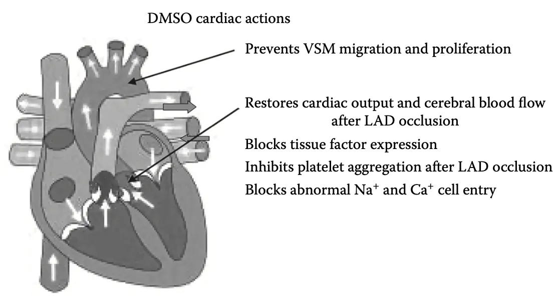
Ischemia and Heart Attacks
As DMSO’s therapeutic properties in strokes directly translate to heart attacks, many studies have explored it here:
•When the heart’s blood supply is impaired, its ability to pump is as well (which is part of why heart attacks are a downhill spiral, as the heart loses the ability to give itself the blood it needs). When heart attacks were modeled (e.g., by ligating the coronary artery to impair the heart’s blood supply), DMSO was able to counteract this. For example, in a dog heart attack study, an IV bolus of DMSO raised cardiac output to 1.65 L/min by the third hour versus 1.15 L/min in untreated controls, alongside lowering systemic vascular resistance and increasing cerebral blood flow.1 Likewise, in another dog study, intravenous DMSO (2 g/kg as a 50% solution*) transiently raised cardiac index from 4.56 to 6.91 L/min/m² and stroke index from 36 to 45 ml/beat/m² while dropping systemic vascular resistance.1
•In addition to preserving the heart’s vital function, many studies have shown DMSO prevents damage to the heart itself. In isolated rat hearts subjected to global ischemia-reperfusion, three days of intraperitoneal DMSO pretreatment (550 mg/kg*) cut infarct size from 50.0% to 21.2% and improved post-ischemic left-ventricular function.1 In rats where the coronary supply was cut for 30 minutes and then reperfused for 120 minutes, DMSO preserved coronary blood flow and reduced both the resulting necrosis (tissue death) by 57.6% and the ensuing left-ventricular dysfunction (an effect that was greater when DMSO was given for several days beforehand rather than immediately prior to occlusion).1 In another rat study, DMSO reduced the damaged portion of the heart by 76.18%,1 and in another it prevented contracture bands from forming in ischemic heart tissue (a common long-term complication of heart attacks).1
•Similarly, in pigs, DMSO significantly reduced intramyocardial hemorrhages and gross pathological changes,1 and in another study improved overall heart attack outcomes.1 In rabbits, giving hydrogen peroxide (H₂O₂) concurrently with DMSO immediately after occlusion reduced the damage still further, presumably by supplying oxygen to the starved tissue.1 Consistent with this, in models of heart attack DMSO reduced the oxidative markers and preserved the antioxidant enzymes that track myocardial injury.1,2
Furthermore, a large part of the damage in a heart attack comes not from the loss of blood flow itself but from its restoration, and DMSO addresses this reperfusion injury directly. In perfused rat hearts, 10% DMSO attenuated the oxygen paradox, reducing reoxygenation creatine-kinase release from 25.3 to 7.2 IU/g and cutting calcium-paradox enzyme release from 134.1 to 54.9 IU/g,1 and it likewise protected against the reperfusion injury caused by calcium-containing solutions.1 In isolated cardiac muscle it preserved contractility during hypoxia,1 an effect echoed in rabbit studies of preserved contractile strength1 and maintained cardiac output and brain perfusion in dogs.1
This same protection extends to hearts held for surgery or storage: in isolated rat hearts, 0.5% DMSO reduced post-storage contractile dysfunction during hypothermic ischemic storage,1 and when added to cardioplegic solutions (used to stop the heart during surgery) at 0.55%, fully preserved mitochondrial respiration after two hours of global ischemia.1 Combined with cardioplegia and alpha-tocopherol,⬖ DMSO likewise improved rat-heart function through ischemia.1,2 Beyond ischemia, it protected against endotoxin-induced myocardial injury,1 improved survival in endotoxemic rats,1 and shielded the heart from intravenous potassium (given at the dose used in lethal injections).1 Most notably, with a cardiac enzyme that normally breaks down ATP (extracted from cow hearts) once mixed with DMSO, it instead synthesized ATP, suggesting DMSO can create a cellular environment that favors energy generation—particularly when the conditions cells normally rely on for it are absent (e.g., during a heart attack).1
Reviews have accordingly proposed DMSO as an antioxidant therapy for myocardial ischemia reperfusion injury1 and summarized its cardiac and CNS pharmacology,1 and reviews of renal and organ protection in aortic surgery note that low-dose IV DMSO restored cardiac output after coronary ligation in dogs while scavenging free radicals, blocking Na⁺/Ca²⁺ influx, and suppressing platelet aggregation.1
Note: as DMSO’s effects are dose and administration dependent, some studies found weaker results. For example, where DMSO served only as the solvent control, it sometimes protected on its own: in a rat deep-hypothermic-circulatory-arrest model testing ebselen, the DMSO solvent-control group itself showed partial protection on several markers, limiting attribution of the benefit to the drug.1 Likewise, in a canine model of coronary occlusion and reperfusion, intravenous DMSO reduced infarct size but not to a statistically significant degree (43.7% vs. 53.4% in controls),1 in two others the benefit was small1,2 and in another heart attack study I located, DMSO provided no benefit (which was likely due to 50% DMSO being directly infused).1 All of this indicates that while helpful, it is not guaranteed DMSO will be able to help every heart attack.
Lastly according to one of the main DMSO authors,1 DMSO was used to treat angina pectoris, heart attacks and prevent heart attacks by physicians in Chile (likely at the dose of 2g/kg). However, despite my best efforts, I could not locate the published paper he referred to with the limited information provided, so it’s likely impossible to corroborate this claim.
Cardioprotective Combinations
Many agents delivered in DMSO reduced infarct size, apoptosis, or oxidative injury in myocardial ischemia-reperfusion and cardiac-arrest models.
Polyphenols & flavonoids — curcumin⬖ (reduced inflammatory cytokines and oxidative stress) 1,2,3,4,5; resveratrol⬖ (reduced oxidative stress and inflammatory cytokines) 1,2,3,4; quercetin⬖ (shrank the infarct) 1; lycopene⬖ (reduced heart-cell death) 1; propolis⬖ (improved heart function) 1; dihydromyricetin⬖ (reduced inflammatory cytokines and cell death) 1; eriocitrin⬖ (improved cell survival and oxidative stress) 1; danshensu⬖ (reduced oxidative stress and cell death); Licochalcone A⬖; curcumin⬖; quercetin (with Ala-Gln)⬖; eriocitrin⬖; tanshinone IIA (PLGA nanoparticles)⬖ (protected against oxidative damage); genistein⬖ (reduced oxidative stress); flavonols⬖ (preserved the heart’s pumping strength); mangiferin⬖ (antioxidant protection against a chemically induced heart attack) 1.
Alkaloids & terpenes — carvacrol⬖ (reduced infarct size and cell death, and reduced mitochondrial fission)1,2,3; higenamine⬖ (reduced infarct size and improved heart function) 1; notopterol⬖ (improved heart function and reduced scarring)1.
Saponins, terpenoids & other botanicals — ginsenoside Rh2⬖ (reduced inflammatory cytokines) 1; ginsenoside 20(S)-Rh2⬖ (improved heart function)1; ginsenoside Rd⬖ (cut infarct size from 36% to 21%, and shifted immune cells toward healing)1,2; magnolol⬖ (reduced infarct size)1; honokiol⬖ (reduced infarct size) 1; asiatic acid⬖ (reduced cell death and oxidative stress)1; curculigoside⬖ (improved cell survival and reduced infarct size)1,2; tanshinone IIA⬖ (reduced cell death and inflammation)1,2; astragaloside IV⬖ (promoted new blood-vessel growth)1,2; triptolide⬖ (reduced inflammation and scarring)1.
Anesthetic (pre-/postconditioning) agents — propofol (reduced cell death)1,2; sufentanil1; sevoflurane (reduced infarct size 36% vs 56%)1,2; desflurane (reduced infarct size 48% to 19%) 1; isoflurane1.
Mitochondrial & metabolic-pathway agents — hydrogen sulfide donors (repeatedly reduced infarct size, including in diabetic hearts)1,2,3,4,5; rapamycin (protected diabetic and normal hearts, cutting infarct size from 36% to 13%)1,2,3,4; Mdivi-1 (reduced infarct size and cell death, including in diabetic and pre-diabetic hearts)1,2; MOTS-c (reduced infarct size)1; diazoxide (reduced oxidative stress and preserved mitochondria)1,2; carbon monoxide-releasing molecules (reduced injury)1,2,3.
Repurposed clinical drugs & metabolic agents — dapagliflozin (reduced inflammatory infiltration)1; canagliflozin (improved heart function)1; lovastatin1; simvastatin (reduced infarct size 39%)1; atorvastatin (reduced infarct size)1; roxadustat (reduced infarct size and cell death)1; ciglitazone (reduced infarct size and inflammation)1; tert-butylhydroquinone (tBHQ)⬖ (reduced oxidative stress); compound C⬖; γ-secretase inhibitor⬖ (reduced oxidative stress); nifedipine and diltiazem⬖ (reduced inflammation and oxidative stress); pioglitazone⬖; NS398 / Ca-channel blockers⬖; sorafenib (toxicity model)⬖; probucol⬖ (reduced ferroptosis, a form of iron-driven cell death); allopurinol⬖; DMOG (HIF-1α activator)⬖; pioglitazone (PA/high-glucose)⬖; agent (equal-volume ip DMSO vehicle)⬖; netrin-1⬖ (reduced oxidative stress and cell death); ferrostatin-1 (reduced ferroptosis)1; 3-iodothyronamine (reduced infarct size)1.
Hormones — 17β-estradiol (reduced microvascular damage)1,2,3.
Cell-death & protease/lipoxygenase inhibitors — ilomastat (reduced infarct size)1; MI-2 (restored the cell’s ferroptosis defenses)1; necrosulfonamide (improved recovery after cardiac arrest by blocking programmed cell death)1; ML355 (improved heart function in pigs and monkeys)1; zVAD-FMK (reduced injury) 1.
Receptor, kinase & other targeted agents — MMI-0100 (cut the loss of heart function by half and halved scarring)1; MHBFC (reduced infarct size and inflammatory cytokines)1; ginkgetin (reduced infarct size and inflammation)1; IB-MECA (reduced infarct size by about 40% in dogs)1; 9-phenanthrol (reduced infarct size from 38% to 9%)1; LGK-974 (improved heart function 62% vs 41%)1; ticagrelor (reduced inflammation)1; MG132 1; L41 (reduced infarct size and spurred heart-muscle regrowth)1; SEW2871 (shifted immune cells toward healing)1; left-ventricular unloading1; HU-210 (reduced infarct size).1
Reader Heart Attack Reports
While CPR can produce miracles, the reality is that it typically doesn’t (e.g., 10.5% of people with heart attacks resuscitated out of the hospital by EMS survive1 and 22-24% of those resuscitated in the hospital survive1). Because of this, I’ve put quite a bit of thought over the years into how this can be improved, and in parallel, we’ve collectively had quite a few cases where something we expected to work (e.g., a natural agent which improves circulation or eliminates clotting) did, including a few cases with DMSO. Given DMSO’s properties described above, I’ve hence repeatedly wondered if injecting DMSO during a cardiac code (CPR protocol) could have saved the person—but due to how unorthodox this was, this was understandably impossible to test. Likewise, while I know of cases where DMSO has been used to treat a heart attack, the reality of the situations around heart attack make giving it quite difficult, so I have much less experience with this than DMSO for strokes. As such, while I suspect it would be lifesaving, that is nothing more than a guess without adequate clinical experience to begin corroborating it and hence not something I can stand behind.
For that reason, I did not previously broach this, but nonetheless a few reader tried it anyways and reported success in stopping heart attacks,1,2,3,4,5,6,7 along with two who stated it healed complications of COVID vaccine induced heart attacks.1,2
Of these, the most interesting one that came from a woman who described her husband getting through what she characterized as a heart attack with a blockage and ischemia, using DMSO and aspirin at the onset and then frequent DMSO over the following days, and coming through it with no measurable heart damage and feeling nearly normal a week later.1 Notably, she independently described the same three-stage structure this section lays out: an initial circulatory phase, in which she believed the DMSO and aspirin opened the blockage in the first minutes; then the oxidative-stress phase of the first few days, in which frequent DMSO was aimed at preserving the heart tissue that reperfusion would otherwise damage; and finally the fibrosis phase from roughly days three to seven, each of which she noted, called for a different approach.1
Note: one reader also reported DMSO helping angina,1,2 and another concerning chest pain.1
Myocardial Necrosis and Fibrosis
DMSO also limits the death and scarring of heart muscle. In rats given isoproterenol (a standard chemical model of infarct-like injury), DMSO reduced fiber necrosis, prevented ventricular aneurysm formation and cardiac rupture, and left a smaller residual area of fibrosis,1 an effect confirmed in a second isoproterenol study,1 in further work showing reduced oxidative stress after isoprenaline,1 and in dedicated work on DMSO and experimental myocardial necrosis.1 Finally, DMSO reduced the several-fold rise in creatine kinase, LDH, and α-hydroxybutyrate dehydrogenase and reduced histologic damage after isoprenaline.1
In a D-galactose accelerated-aging model, subcutaneous 4% DMSO lowered TGF-β1, normalized cardiomyocyte diameter, and reduced fibrotic area to near-control levels, an anti-fibrotic effect the authors attributed to DMSO itself.1 In a series of studies on copper deficiency (which drives cardiac hypertrophy), chronically administered DMSO inhibited the hypertrophy, anemia, and heart-copper depletion via hydroxyl-radical scavenging, attenuated the rise in heart weight and water content, and preserved mitochondrial and myofibrillar ultrastructure.1,2,3 Consistent with a radical-mediated mechanism, DMSO also reversed the copper-induced impairment of force generation, calcium handling, and myosin-ATPase activity in cardiac muscle1,2
Note: DMSO also protected older rat hearts from the stress caused by prolonged immobility and from dietary copper deficiencies.1,2,3,4,5
Cardiac Fibrosis & Remodeling Combinations
In the fibrosis and remodeling literature, the same anti-fibrotic or anti-hypertrophic effects was seen with many agents delivered in DMSO.
•Natural agents — curcumin⬖ (raised antioxidant defenses and reduced heart-muscle damage); curcumin⬖ (protected against heart-muscle damage); genistein⬖ (reduced hypertrophy, oxidative stress, and inflammation); atractylenolide III⬖ (reduced post-heart-attack scarring); gCTRP9⬖ (protected injured heart cells); RGS6 with DPI⬖ (reduced remodeling); CRIF1 with NAC⬖ (protected injured heart cells); Maresin 1⬖ (protected heart cells); andrographolide⬖ (reduced hypertrophy and scarring); baicalein⬖ (reduced heart-muscle damage, scarring, and inflammation); β-carotene⬖ (reduced inflammation and scarring); resveratrol⬖ (reduced fibroblast overgrowth); tanshinone IIA⬖ (reduced hypertrophy); asiaticoside⬖ (reduced hypertrophy); salvianolic acid B⬖ (improved heart function); anacardic acid⬖ (blocked hypertrophy); colchicine⬖ (reduced fibrosis); nimbolide⬖ (reduced fibrosis in diabetic hearts); Buyang Huanwu Decoction⬖ (reduced post-heart-attack cell death).
•Pharmaceutical & synthetic agents — valsartan (reversed chemotherapy-induced remodeling)1; a JNK inhibitor (reduced fibrosis)1; a LOXL2 inhibitor (reduced fibrosis and hypertrophy)1; an EZH2 inhibitor (reduced fibrotic proteins)1; a GCN5 inhibitor (reduced hypertrophy and fibrosis)1; salubrinal (improved function in heart failure)1; rapamycin (improved pressure-overload hypertrophy)1; a Pannexin1 inhibitor (reduced hypertrophy and fibroblast activation)1; an H2S donor (reduced remodeling and blood pressure)1; dasatinib (reduced fat buildup and fibrosis in diabetic hearts)1.
Cardiotoxicity, Sepsis, and the Diabetic Heart
In 42 severely ill patients with septic complications of post-resuscitation disease, intravenous DMSO was an effective therapy, including in cases where the sepsis arose from antibiotic-resistant bacteria.1
Additionally, DMSO has been used with a wide variety of agents to protect chemically injured, septic, and metabolically stressed hearts where it frequently reduced cardiac enzymes, oxidative stress, inflammation, and apoptosis:
•Chemotherapy cardiotoxicity — Cissus verticillata⬖; Irvingia gabonensis extracts⬖ (reduced heart-injury markers); ursolic acid⬖1,2 (improved heart function and reduced cell death); silibinin⬖ (promoted protective autophagy); necrosulfonamide (restored antioxidant enzymes)1; necrostatin-1 (reduced heart-injury markers and inflammation)1; a PARP inhibitor (reversed apoptosis and remodeling)1; azilsartan (reduced oxidative stress and cell death)1,2; selenium⬖ (reduced inflammation and cell death); avenanthramide-C⬖ (reduced oxidative stress and inflammation); rutin⬖ (raised antioxidant defenses and reduced inflammation).
•Sepsis-induced cardiac injury — Trimetazidine⬖ (protected against sepsis heart injury); D3T⬖ (raised antioxidant defenses); a pancreatitis-ascitic-fluid injury model (DMSO solvent control); paeoniflorin⬖ (reduced vascular leakage and inflammation); astragaloside IV⬖ (reduced oxidative stress and inflammation); astaxanthin⬖ (reduced injury signaling); curcumin⬖ (reduced heart-injury markers); luteolin⬖ (enhanced protective autophagy); sulforaphane⬖ (reduced inflammation); xanthohumol⬖ (reduced inflammation and cell death); octreotide (reduced inflammation and raised antioxidant defenses)1; simvastatin (reduced heart-injury markers and inflammation)1.
•Diabetic cardiomyopathy — GLP-1 drugs⬖ (reduced oxidative stress in heart blood vessels); tadalafil (protected mitochondria)1; rapamycin (reduced oxidative stress)1,2; ferrostatin-1 (reversed ferroptosis, a form of iron-driven cell death)1; honokiol⬖ (reduced high-glucose fibrosis); resveratrol⬖ (reduced inflammation); EGCG⬖ (restored cell-to-cell coupling).
Combinations for Additional Heart Conditions
In pulmonary hypertension and pulmonary embolism, where the right heart is strained by elevated pressures, DMSO delivered agents benefited right-ventricular function and pulmonary vascular remodeling:
•Myocarditis — A proteasome inhibitor (reduced inflammation and apoptosis);1 an α7-nicotinic agonist,1,2 (reduced inflammation and cell death); carbon monoxide-releasing molecules (reduced mortality and heart injury);1 a 3C protease inhibitor (antiviral protection);1 an androgen-receptor inhibitor (reduced inflammation and enhanced autophagy);1 a tyrphostin (reduced inflammation).1
•Arrhythmias — A calpain inhibitor (reduced atrial-fibrillation incidence); mexiletine (shortened repolarization in long-QT models); fisetin⬖ (reduced atrial inflammation and fibrosis after heart attack); valsartan or an ERK inhibitor (reduced atrial fibrosis and remodeling).
•Pulmonary Hypertension (which also damages the heart)—Curcumin⬖1,2 (reduced platelet activation and heart-injury markers); resveratrol⬖ (reduced inflammation and heart strain); isoliquiritigenin⬖ (reduced right-heart pressure and oxidative stress); icariin⬖ (reduced inflammation); a fission inhibitor with 17β-estradiol (reduced right-heart enlargement and shifted immune cells toward healing); a TRAF6 inhibitor (improved right-heart function); a Pannexin-1 inhibitor (improved pulmonary artery flow).
Direct Effects on Heart Function
Several of DMSO’s standalone cardiac actions cut across all of the above. DMSO is a direct vasomotor agent, producing endothelium-independent relaxation of coronary and pulmonary arteries1. As a hydroxyl-radical scavenger it also attenuated tempol-induced falls in blood pressure, heart rate, and sympathetic nerve activity1. As discussed earlier, DMSO also protects coronary endothelium-dependent relaxation against oxidative injury, inhibits platelet aggregation, and suppresses tissue-factor expression and arterial thrombus formation—vascular protections equally relevant to the heart. A DMSO–aspirin preparation was likewise reported to reduce erythrocyte aggregation, normalize fibrin formation, lower peripheral vascular resistance, and improve microcirculation1. In the vehicle-delivered atherosclerosis and vascular-injury literature the same effects recur, with cinnamaldehyde⬖1 cutting serum lipids and raising heart antioxidant enzymes in atherosclerotic rats, hesperetin⬖1 limiting diesel-exhaust cardiovascular oxidative damage, and kolaviron⬖1 attenuating angiotensin-II/LPS-driven vascular smooth-muscle proliferation.
On the muscle itself it exerts direct, concentration-dependent effects on contractility, rate, and electrophysiology1,2,3: at low doses positive inotropy1—a moderate increase at 3% becoming marked at 6%1—while potentiating the inotropic response to isoprenaline1; still higher concentrations reversibly depress contractility1 or produce a mild hyperpolarization that prolongs the action potential1. These effects are independent of beta-adrenergic receptors1 and typically do not alter cardiac rhythm1, though the rate response is dose-dependent: 1–3% DMSO slightly raised heart rate while 6–10% markedly slowed it, an effect reversed by atropine1 and thus mediated through cholinesterase inhibition.
Note: DMSO and colchicine were found to increase myocardial work output.1
DMSO increased cardiac output during hypoxia1 and, given IV to dogs, raised cardiac output, stroke volume, and central venous pressure1. DMSO further protects cardiac t-tubules from stress-induced sealing1. On the parasympathetic side it inhibits cardiac acetylcholinesterase, lowering the vagal threshold and potentiating acetylcholine’s slowing of the heart rate.1
As discussed earlier, DMSO’s radical scavenging protects cardiac calcium handling, the sodium-potassium pump, and heart-muscle contractility against iron- and peroxide-driven injury making antioxidant protection equally relevant here. Beyond those, in cell-free and isolated-heart systems DMSO acts as a direct radical scavenger on its own: it neutralized electrolytically generated oxygen radicals in an enzyme-free buffer1, protected rabbit hearts against radical-driven rises in coronary and end-diastolic pressure and albumin leakage1, raised coronary release of protective uric acid in guinea-pig hearts1, and served as the reactive-oxygen scavenger confirming the antioxidant basis of protection against ischemia-reperfusion injury1. It also limits stress-induced lipid peroxidation in the myocardium1 (most pronounced in aged hearts), and in H9c2 cardiomyocytes it upregulates heme oxygenase-1 via p38 MAPK and Nrf2 to protect against hydrogen-peroxide injury1.
Differentiation of Heart Cells
DMSO has long been used to turn cancer cells back into normal cells and to steer stem cells into the cell types the body needs — a way to replace and repair damaged tissue, including the nervous system. A large body of work shows the same thing for the heart: DMSO alone reliably drives stem cells to become cardiomyocytes.
When stem cells are exposed to DMSO alone they are most often reported to become heart tissue, which points to a real role for DMSO in cardiac stem-cell protocols.
A very large literature has since used DMSO as a cardiomyocyte-differentiation inducer across many starting cell types, with the same result recurring: exposure to DMSO (typically 0.5 to 1 percent) drove the cells to switch on cardiac genes and structural muscle proteins and, in most reports, to form spontaneously beating cardiomyocytes.
By far the most studied model is the P19 (and P19CL6) embryonal carcinoma line, in which DMSO alone reliably produced beating cardiomyocytes and became the standard system for dissecting how it works.t1,2,3,4,5,6,7,8,9,10, 11,12,13,14,15,16,17,18,19,20, 21,22,23,24,25,26,27,28,29,30, 31,32,33,34,35,36,37,38,39,40, 41,42,43,44,45,46,47,48,49,50, 51,52,53,54,55,56,57,58,59,60, 61,62,63,64,65,66,67,68,69,70, 71,72,73,74,75,76,77,78,79,80, 81.
The effect shows up across source cell type after source cell type. DMSO alone has turned bone-marrow mesenchymal stem cells,2 into myocardial cells, adipose-derived stem cells into cardiomyoblast-like cells, and fetal liver stem cells into cardiomyocyte-like cells. It has also driven fetal cardiomyocytes to proliferate and boosted the direct reprogramming of fibroblasts into cardiomyocytes roughly five-fold.
The same DMSO-driven differentiation has been reported across species and stem-cell source: in goat embryonic stem cells, mouse embryonic stem cells1,2,3,4,5, human embryonic stem cells1,2,3,4,5, human bone-marrow stem cells, cardiac progenitor cells, and mouse embryonic fibroblasts.
Lastly, the same differentiation was also achieved when DMSO was combined with many other agents including 5-azacytidine, a cocktail of growth factors including BMP and FGF, retinoic acid with 5-azacytidine, oxytocin (which worked synergistically with DMSO) and many others.1,2
Note: the effect is again concentration-dependent. At high concentration the picture reverses: a brief exposure to 10 percent DMSO disrupted muscle fibers and suppressed proliferation in cultured heart and cardiac endothelial cells,1,2
Atherosclerosis
DMSO endothelial protective effects also allow it to prevent atherosclerosis itself. In rabbits fed a cholesterol-overloaded diet, oral DMSO reduced the resulting atherosclerosis by 30—40% and halved the cholesterol accumulating in the tissue.1 Beyond limiting the damage, it also supported the vasculature’s ability to rebuild itself, as DMSO promoted the differentiation of human embryonic stem cells into endothelial, cardiac, and blood-cell precursors—the early cells the body draws upon to build new vessels and blood.1
Finally, a series of studies in the cells lining human umbilical veins (a standard laboratory stand-in for the vessel wall) point to a protective and anti-inflammatory action there. Pretreatment with 2.5% DMSO reduced the neutrophil adhesion TNF-α normally triggers, which is one of the first steps of vascular inflammation and the initiating event in atherosclerosis.1 In nutrient-starved cells it blocked programmed cell death by promoting DNA replication and enhancing survival,1 and under oxidative stress it raised heme oxygenase-1 and reduced apoptosis through several anti-inflammatory and cytoprotective pathways.1
Blood Pressure
As DMSO is a mild vasodilator which simultaneously increases parasympathetic tone and scavenges the radicals that stiffen and constrict vessels, there is a mechanistic basis for it reducing blood pressure. However, the direct evidence is quite limited and most of what exists is indirect.
For example, DMSO is used as a transdermal penetration enhancer for conventional antihypertensives such as diltiazem and labetalol, where it improves both how much of the drug crosses the skin and how far the pressure falls in hypertensive animals.1,2 More importantly, it protects the vessels hypertension damages rather than merely the number on the cuff, as in rats made hypertensive with angiotensin II, DMSO reduced vascular hyperpermeability, smooth-muscle necrosis, and fibrin deposition in the small arteries by scavenging free radicals1—guarding the vessel wall even in cases where it did not move the pressure at all.
Note: as I’ve shown throughout the series, the primary value of DMSO for cardiovascular disease is its ability to protect the tissue rather than meet a numerical blood pressure threshold (which is often not even healthy).
DMSO is not, however, a blood pressure drug in the way an ACE inhibitor is, and as with every other system, its effect is concentration dependent. At the low solvent-level doses used in most research it is close to neutral, as DMSO-alone control groups repeatedly show no meaningful change in mean arterial pressure.1,2 Any lowering emerges only at the higher therapeutic concentrations where the vasodilation becomes pronounced, and at the far end of the range the direction reverses entirely—the same counter-current already seen when DMSO scavenged enough nitric oxide to nudge blood pressure up in horses. The clearest illustration of this is what happens when stem cell grafts cryopreserved in concentrated (10%) DMSO are infused rapidly, which produces a transient rise in blood pressure proportional to the DMSO dose1—the same high-concentration ceiling described in the safety section above. In short, DMSO is a mild and dose-dependent vasodilator whose most consistent contribution in hypertension is protecting the blood vessels rather than driving down the number on the cuff.
Readers, in turn, have described exactly what the mechanism predicts, and frequently stumbled upon it by accident. One woman had been applying DMSO to her husband’s belly at night for a pulled muscle when a checkup found his blood pressure had normalized; months of magnesium and potassium had only brought him to 150/90, but he now read 125/80.1
The pattern which recurs the most however is a rapid drop following a single oral dose. A 73-year-old found half a teaspoon in water took him from a usual 148/90 to 109/70 within thirty minutes, which he described as “way better than the meds I used to take.”1 Another, who had been pushed up to 40 mg of lisinopril and was applying DMSO over his carotids, watched a pre-DMSO 159/95 fall to 106/75 on the first reading afterwards.1 Others reported oral DMSO doing “more for my general inflammation and BP than aspirin or these days BP meds,”1 blood pressure finally dropping after years of unsuccessful diet, exercise, and weight changes,1 and a more modest shift from 160/90 to 150/80 which arrived alongside easier urination and sharper vision.1
Consistent with this being vasodilation rather than something else, a few readers instead noticed it as mild low-pressure symptoms when they used DMSO aggressively, one describing feeling “rather unsteady” with unusually flat and invisible hand veins during heavy use1 (which the German DMSO community believes accounts for why some people get temporary headaches from significant DMSO use). In others it was simply folded into a much broader recovery, such as the reader treating a vaccine injury with oral DMSO who listed normalized blood pressure among a long list of improvements,1 and another who found his blood pressure and cholesterol returned to normal once DMSO allowed him to discontinue Humira.1
Given all of this, I would expect DMSO to help certain kinds of blood pressure but not others (as many different things can cause the elevation). That said, blood pressure is not always a thing you want to treat (sometimes it being elevated is helpful)—although in the cases where it’s helpful to be elevated (to maintain perfusion through aging arteries), DMSO would likely at least somewhat bridge the gap due to its ability to promote circulation.
Note: because a few of these drops were both large and rapid, anyone already on blood pressure medication should monitor their pressure when starting DMSO rather than assume the two simply stack, as the reader above who was on 40 mg of lisinopril illustrates how quickly the combination can move things.
Peripheral Circulatory Disorders
In addition to protecting tissues from death, DMSO is remarkably effective at removing excess fluid from outside the bloodstream, increasing circulation, and clearing circulatory obstructions such as clots, along with many other conditions which result from impaired circulation (and microcirculation).
Note: numerous observations of the years have led me to believe blood congestion is a primary cause of many ailments including varicose veins and hemorrhoids.
The pioneering DMSO researchers documented this early. Stanley Jacob found1 that half of Raynaud’s patients had their symptoms eliminated and that thrombophlebitis responded excellently, while two investigators using plethysmography demonstrated objective improvement1 in peripheral-artery insufficiency in a large number of patients given topical DMSO. DMSO was also shown to relieve diabetic circulatory problems including peripheral neuropathy1 and diabetic ulcers, where one study1 of hundreds of patients reported over a 94% success rate — and to help prevent future amputations (data which has since been extensively corroborated).
The early clinical series were substantial. A study of 67 patients with varicose ulcers1 (39 women, 28 men) found a remarkable response even in chronic ulcers that had been present for years and had failed other treatments. Likewise a larger early1 study found:

DMSO’s vasodilatory action has long been invoked for frostbite, diabetic ulcers, and varicose ulcers1, and over a hundred studies have since corroborated these results.
Venous disease and varicose veins. The most consistent modern use is topical, much of it centered on Dolobene — a gel of DMSO, heparin, and dexpanthenol in which DMSO both carries the heparin through the skin and adds its own analgesic and anti-inflammatory action. Dolobene has been evaluated across the venous disorders1, used specifically for varicose vein disease1, and ranked by vascular surgeons and pharmacists among the priority topical agents for varicose veins1.2,3,4,5 An early double-blind study of 55 patients1 tested topical DMSO for chronic venous insufficiency, and a 1975 New York Academy of Sciences report1 documented its use across venous stasis, dermatosclerosis, and lymphedema. It recurs throughout the topical treatment of chronic venous insufficiency and post-thrombophlebitic disease1 and appears in the formulation literature for antivaricose gels1 and horse-chestnut/aescin preparations1 as the penetration enhancer of choice.1 This indicates DMSO may also play a wall-strengthening role (e.g., in a mouse model of venous hypertension1 that reproduces varicose remodeling, agents delivered transdermally in DMSO prevented the pathological changes in vein structure and smooth muscle that drive the disease).
Thrombophlebitis, superficial venous thrombosis and hematomas. DMSO-based gels are commonly used here. A randomized, double-blind trial of a Phlebolan spray1 and related studies1,2,3 support topical DMSO-heparin (and the topical DMSO heparin gel Dolobene) for superficial thrombophlebitis.1 Likewise Dolobene recurs as a standard thrombophlebitis and thrombosis therapy throughout the literature, including for combined deep and saphenous-vein thrombosis.1,2,3 DMSO (alone) gels have also been used to treat acute thrombophlebitis1, superficial-vein thrombosis1, and chemotherapy-induced phlebitis.1,2,3 Electrophoretic DMSO has also resolved intramuscular-injection hematomas in hemophilic children, clearing the bruising well enough to let their therapy continue.1
Venous, trophic, and diabetic ulcers. DMSO’s ulcer record (e.g., the greater than 94% success rate and the 67-patient varicose-ulcer series above) are highlighted in many papers discussing the use of large DMSO containing wound-dressing for venous trophic ulcers and lipodermatosclerosis1, chronic trophic leg ulcers1, post-thrombophlebitic trophic ulcers1, venous ulcers and chronic venous insufficiency (in one study a DMSO powder healed 95% of patients at 3 months versus 70% on placebo),1,2 and trophic ulcers of atherosclerotic origin (with a DMSO powder)1. In 58 patients with chronic purulent wounds from vascular disease or diabetic angiopathy, negative-pressure instillation of 20% DMSO produced faster cleansing, near-complete granulation in 76% by the first dressing change, and healing 1.5–3× faster than standard care.1,2 The diabetic-foot thread is distinct: in 80 diabetic patients with lower-extremity ulcers, DMSO dressings combined with an oxygen-carrying perfluorocarbon cut mean healing time to 17 days versus 47 for standard care, with faster bacterial clearance and a better microcirculatory index by day 22.1 DMSO is also used as an antiseptic and penetration enhancer in diabetic-foot and gangrenous-wound protocols1, in diabetic wound management with documented perfusion improvement1, for necrobiosis lipoidica and other diabetic skin conditions1, and as the vehicle in experimental diabetic-wound and angiogenesis models.1
Scleroderma and Raynaud’s. In addition to the 50% Raynaud’s symptom-elimination figure from the original DMSO literature, DMSO is a standard component of scleroderma care specifically for its microcirculatory action. DMSO with nicotinic acid⬖ has been used to treat systemic scleroderma,1 and topical DMSO (often paired with heparin) is mentioned as a treatment throughout the scleroderma treatment literature1, including pediatric localized scleroderma1, explicitly to improve microcirculation and soften dermatosclerosis.1,2,3,4,5,6,7,8 (where in one study, blood flow was increased 20% by DMSO and 500% by DMSO with nicotinic acid⬖1). It also appears in systemic lupus with acrocyanosis and Raynaud-type lesions1, in vasculitis1 with cutaneous circulatory involvement, and as the vehicle for vasoactive agents in scleroderma1.2
Note: many more scleroderma studies are discussed here.
Tissue perfusion, flap and graft survival. The most objective experimental evidence that DMSO raises tissue blood flow comes from surgical flap models, where perfusion decides whether ischemic tissue lives. In 48 rats with abdominal island flaps1, systemic DMSO significantly increased perfusion (by laser Doppler and perfusion fluorometry) and improved flap survival. In ischemic dorsal skin flaps1, 5% DMSO reduced distal necrosis and produced greater neovascularization and earlier repair. The benefit recurs across independent models such as pedicle-flap necrosis1,2,3,4 composite and vascular allografts1, or nipple-areolar and penile tissue salvage1.2 The same perfusion gain runs through a large wound-healing literature, where DMSO (alone or as carrier) increases skin blood flow1, normalizes capillary permeability and microcirculation1, improves isolated-organ perfusion1, and accelerates granulation and revascularization in trophic ulcers, burns, and other wounds.1,2,3
Note: many additional DMSO skin flap studies can be read here.
Surgery. In addition to protecting skin flaps (a common challenge in plastic surgery), DMSO also addresses many other challenges. In breast reconstruction, applying topical 60% DMSO before each tissue-expansion session cut expansion time by roughly a third, allowed greater volume per session, and reduced pain1, across two clinical series.1 Topical DMSO salvaged (saved) an ischemic nipple-areola complex1 after reduction mammoplasty by restoring perfusion, and as a trauma wound dressing used in nearly ten thousand cases it dilated capillaries and improved microcirculation1. Finally, in 154 patients undergoing surgery to restore blood flow to an acutely ischemic leg, intravenous DMSO added before reperfusion reduced markers of reperfusion injury1 ( lower peak lactate, faster normalization, fewer cases of kidney and cardiopulmonary dysfunction) though amputation and mortality were unchanged.
Hemorrhoids and frostbite. DMSO is used in topical mixtures for acute thrombosed hemorrhoids1 and hemorrhoidal thrombosis1, in a placebo-controlled trial of an MSM⬖-containing gel1 (MSM being DMSO’s oxidized metabolite), and in anorectal wound healing after hemorrhoid surgery1.2,3 For frostbite, a peripheral cold-ischemic injury, DMSO appears in the treatment guidelines for local cold trauma1, in military cold-injury research1, and as a carrier delivering heparin into cold-injured tissue1.2,3,4,5
Note: Stanley Jacob (the pioneer of DMSO) treated hemorrhoids with DMSO,1 other DMSO authors have advocated for using DMSO to treat them, and Merck (in their early clinical trials) reported that it improved recovery after their surgical removal.1
Arterial and ischemic limb disease. The arterial side is thinner than the venous but present: DMSO has been used adjunctively for arterial disease of the extremities1, for chronic purulent wounds from advanced peripheral arterial occlusive disease1, with hyaluronidase for ischemic limb edema1, and for vaso-occlusive hand-foot crises1 where Dolobene relieved pain and edema. It also improved facial arterial blood flow after surgery1.
Veterinary peripheral circulation. In veterinary medicine, where DMSO is a mainstream drug, it is used for equine laminitis to improve digital perfusion1, distal-limb edema1, and aorto-iliac thrombosis1, in regional limb perfusion1 and tendinitis1, for bovine hoof and teat lesions1, and as anti-endotoxemic support in colic and peritonitis1 where circulatory collapse is the threat.1,2
DMSO also completely abolished toxin-induced constriction of lymphatic microvessels1, protecting lymphatic as well as blood-vessel tone.
Peripheral-Vascular Combinations
Many agents have also been combined with DMSO to topically treat the same conditions DMSO alone also treats.
Venous Disease, Trophic Ulcers & Wound Healing
Many agents combined with DMSO, like DMSO alone, speed cleansing, granulation, and closure of venous, trophic, diabetic, and traumatic wounds:
•Antiseptics & antibiotics (topical wound care) — furacillin + sensitivity-selected antibiotics (25% DMSO cleared monoflora by day 5–6 and healed varicose/post-thrombophlebitic ulcers in 16 days, 2.2x faster with phototherapy)1; chlorhexidine (multimodal venous trophic-ulcer regimen; remission in 80–95% of 152 cases)1; bactosin + sulbactam (DMSO antiseptic irrigation of contaminated vascular-trauma wounds; fewer thromboses, infections, amputations),1 photoditazine plus photodynamic therapy (gunshot wound treatment aiding cleansing and microcirculation)1,2.
•Anesthetics & anti-inflammatories (compounded dressings/ointments) — Anikol, a patented anilocaine ointment (~190-min surface anesthesia; infected wounds closed in 7 days, frostbite healed in 8)1; splenic preparation splenodimexide (anorectal wounds; complications cut >1.5x, faster granulation)1
•Free-radical scavengers & flavonoids (ulcer healing) — Quercetin⬖ (10% DMSO vehicle showed no independent effect while quercetin-in-DMSO drove angiogenesis and closure in diabetic wounds)1; loureirin A⬖ (promoted vascularization and wound healing via miR-339-5p/Wnt).1
Thrombosis, Clotting, Platelet Function & Vascular Tone
DMSO is frequently combined with other anti-clotting agents:
•Natural compounds — curcumin⬖ (dose-dependently reduced platelet aggregation, CD63, GMP-140 in pulmonary embolism)1; berberine⬖ (prolonged mesenteric-artery occlusion time; reduced venous thrombosis and NET formation in colitis)1; senkyunolide I (reduced platelet activation and NET formation in sepsis).1
•Anticoagulants & other pharmaceuticals — rivaroxaban (suppressed leukocyte adhesion and microthrombus formation in diabetic microvasculature)1; aspirin (DMSO + aspirin stopped nocturnal leg cramps within a day)1. Nadroparin (15% DMSO carried this LMWH into tissue to prevent Nicolau syndrome skin necrosis after sclerotherapy)1; hydrocortisone (90% DMSO potentiated hydrocortisone ~10-fold and, alone, prolonged arteriolar flow-stop time and sped flow restoration after venous thrombosis)1; Procaine + heparin (corrected vascular tone and arterial-bed filling in chronic interstitial parotitis).1
•Signaling-pathway inhibitors (thrombosis mechanism) — PI3K inhibitor wortmannin (malnutrition-driven venous thrombosis)1; suberoylanilide hydroxamic acid(improved survival and clot quality in sepsis coagulopathy)1; Tubastatin A (HDAC6) (reduced renal microcirculatory thrombin and NET formation in sepsis).1
•Polyphenols & natural vasorelaxants — resveratrol⬖ (near-complete relaxation of human intrapulmonary arteries, partly NO-dependent)1; garlic diallyl trisulfide⬖ (endothelium-independent relaxation of intrarenal arteries, Emax >91%)1; tetrahydroxystilbene glucoside⬖ (mesenteric relaxation via COX-2/TXA2 and K+/Ca2+ channels)1; cannabidiol (potent inhibition of small-resistance-artery contraction, stronger in smaller vessels)1; resveratrol ⬖ + CAPE⬖ silibinin⬖ (concentration-dependent relaxation of human umbilical artery).1
Endothelial Protection & Microvascular Barrier
Polyphenols & flavonoids — resveratrol⬖ (raised hypoxic endothelial viability and SIRT1/PGC-1α; lowered inflammatory IL-6, ICAM-1, ROS)1; resveratrol⬖ plus atorvastatin (synergistically enhanced bone marrow stem cell endothelial differentiation and stent re-endothelialization beyond either alone)1; kaempferol⬖ (protected islet microvessel endothelium from fatty-acid injury)1; astragaloside IV⬖ (improved aortic-endothelial viability, migration, tube formation under hypoxia).1
Endothelial-barrier disruption — ulinastatin (blocked endothelial hyperpermeability via p38)1; HSPA12B pathway inhibitors (preserved endothelial barrier function),1 thymoquinone⬖ (sepsis) (improved mesenteric-artery blood flow and aortic function,)1. resveratrol⬖ (cut sepsis leukocyte/platelet adhesion, improved survival),1 genipin⬖ (reduced LPS-induced vascular hyperpermeability via SIRT1).1
Neointimal Vascular Remodeling after Artery Injury — celastrol⬖ (restored contractile VSMC markers)1; ursolic acid⬖ (also reduced NF-κB/PCNA)1; YM2016361; a Pol I inhibitor;1 enalaprilat (DMSO alone demonstrated a modest reduction in remodeling).1
Reader Reports
Since DMSO is easy to utilize for peripheral circulatory conditions, many readers have attempted it, and their testimonials mirror the successes for these conditions seen in the trials.
Varicose and spider veins. This is the single most common circulatory use readers describe, and the reports are strikingly consistent: repeated topical application (typically 70-100% DMSO,* often in aloe⬖ or castor oil,⬖ once or twice daily) shrinks, softens, fades, and in many cases eliminates the veins, frequently after they had been present for decades. One reader sprayed 100%* DMSO on varicose veins nightly and reported that “in 13 months they are entirely gone,”1 another in New Zealand treated badly “varicosed” saphenous veins roughly half a dozen times over a week and found the change (documented in before-and-after photos) durable: “I am fairly sure that the applications are not maintaining the varicose vein recovery-they are fixed.”1 A third watched veins that “stuck out about 1/2 inch” recede to “about 1/8 of an inch,”1 and one posted a 24-hour before-and-after showing a large varicosity essentially gone.1 Many more described veins that “just disappeared” along with the aching and end-of-day leg pain,1 that faded or shrank steadily with continued use1,2,3,4,5,6,7 that became “much smaller, softer” with the previous sensation of “thickness” gone1 or that improved only on the treated leg while the untreated leg served as an inadvertent control.1 A woman treating her elderly relative’s chronic venous insufficiency tracked it week by week—roughly 20% improvement in the first week, 50% by the second, 75% by the third.1 Several noted the veins returned when treatment stopped1 and a minority saw little or no effect,1,2 but the dominant pattern was steady improvement of veins that conventional medicine treats only by stripping or ablation.1,2,3,4,5,6
These are some of the many pictures I’ve received:


Blood clots and DVT. Readers reported both topical and oral DMSO dissolving existing clots and preventing new ones, often as a deliberate replacement for prescribed anticoagulants. One who had been told to take Eliquis for life stopped it and switched entirely to oral DMSO to dissolve clots in the legs and lungs: “I’d be dead if DMSO didn’t work.”1 Another treated a bad DVT running from above the knee to the ankle with topical DMSO (plus lumbrokinase⬖ and serrapeptase⬖) and “dissolved the clot in 2.5 months,” declining the year of Eliquis doctors advised.1 A mother whose son had hip-bone necrosis from a clot lodged in a small twisted vein reported that after starting DMSO “within 3 days he was able to walk without pain,” and that the hip healed without the scheduled surgery.1 One reader whose arm had visible hardened clots from an IV reaction for months found “the clots were gone within three days” of applying DMSO gel,1 and another watched a large hard lump on a calf vein resolve until “it is gone now and the area soft.”1 Another described using DMSO to resolve a clot in an 89-year-old’s leg.1
Raynaud’s and cold extremities. For Raynaud’s, the reported effect is often nearly immediate. One reader who came in from shoveling snow put DMSO on the fingertips and was “instantly warm”;1 another applied it to the toes and it “changed the color of my toes back to normal.”1 A regular user reported applying it two or three times daily to her hands with “great success, no longer needing to wear fingerless gloves around the house...It has been life changing,”1 while another with cold hands happened to track the effect on a pulse oximeter and watched readings climb after each application.1 Others described relief of the spasms, discoloration, and stiffness,1,2,3 marked overall improvement in circulation alongside the Raynaud’s,1 and one reported that five consecutive days of DMSO “cured Chilblains and Raynaud’s for the entire winter season.”1 Several with autoimmune-associated Raynaud’s (lupus, scleroderma, rheumatoid disease) folded it into broader regimens with reported relief.1,2,3
Ischemic and discolored limbs. Many reported normal color and warmth to feet and legs that had gone blue, purple, or black from poor perfusion. A daughter treating her 85-year-old mother’s severe foot neuropathy (toes and ankles “a blackish blue color,” numb, with nightly cramping) described the results after three weeks as “amazing” “the normal color has returned to her feet and legs...she now has feeling back in both of her heels.”1 Another reader rubbing DMSO cream on the feet nightly found that “on the fourth morning I looked down at my feet and all the purple mottling was completely gone.”1 One person watched an elderly mother’s “blue-black toes...from frostbite” return “to a healthy pink—permanently” after a single application of DMSO with magnesium.1 An 84-year-old with a decades-old radiation wound near the bone that had never fully healed reported that once she applied DMSO gel, “the skin color has returned to normal and circulation is now almost normal, my foot does not swell from lack of circulation.”1 Many others described impaired circulation, cold feet, leg swelling, and perfusion-related pain improving with topical or oral use1,2,3,4,5,6 including a post-COVID reader whose cyanotic, “bluish purple” legs looked “a normal healthy colour...for the first time in 3 years.”1
Frostbite and cold injury. Frostbite is a peripheral cold-ischemic injury, and readers reported DMSO restoring warmth and color to affected tissue, often within hours, particularly when applied promptly. One who “burned” a hand with liquid refrigerant until “half of my hand went numb and changed color” applied DMSO and reported the hand “back to normal” two days later, with only a little surface skin peeling.1 A second described the same refrigerant injury (a hand “numb for 24 hours and changing color”) returning to normal in three days, adding that “immediate or even preemptive application is essential.”1 Another reported that on a “almost frost bite,” DMSO “restored warm asap.”1 These mirror the documented reports of frostbitten and blue-black toes regaining healthy color after DMSO.
Venous, diabetic, and pressure ulcers. DMSO’s reported effect on chronic wounds and ulcers is among the most consistent in this collection. A reader watched an elderly friend’s leg “a six inch diameter festering mess” that a year of medical treatment had failed to close, with surgeons wanting to cut “turn into clean skin” over a month of DMSO.1 A 12-year user reported that DMSO “saved a 75-year-old woman’s leg” doctors wanted to amputate: “she walks now despite being bedridden before.”1 Wives caring for diabetic husbands with chronic venous ulcers—one after a year of wound-care visits and twice-weekly home nursing—described the ulcers improving significantly within two weeks of switching to DMSO gel1 and not recurring afterward.1 A diabetic reader healed three foot ulcers with DMSO gel and pursued IV infusions to improve leg blood flow,1 and a man given DMSO cream by an acquaintance saw “a 2 inch ulcer” on his arm heal over “in a couple of weeks.”1 The same pattern appears with pressure sores: a family applied 80% DMSO to a bedbound 101-year-old’s bedsores and reported her skin “became normal in just a matter of days,”1 and a caregiver treating early Alzheimer’s incidentally cleared his wife’s “horrid bed sores,” leaving her “with no sores or irritated pink skin.”1 Others reported healing of diabetic leg ulcers, lupus-related skin ulcers, and assorted chronic sores, often in combination with iodine, silver, or chlorine dioxide.1,2,3,4,5,6
Hemorrhoids. Because a hemorrhoid is essentially a swollen, clotted vein, readers repeatedly found it responded to DMSO the way varicosities did—often within days (to the great surprise of skeptical readers).1 One applied “two drops of DMSO on a hemorrhoid I had for years” and reported that “after a single dose, it shrank to the size of a grain of sand.”1 Another reported that two friends “had their hemorrhoids vanish after a single subcutaneous thigh injection of .5ml of DMSO,” including one who had had them for 15 years.1 A reader whose doctor had diagnosed a hemorrhoid and written a prescription instead applied DMSO to the bulge and reported it “gone” after three days,1 and another who makes DMSO-cocoa butter suppositories reported they “heal and relieve hemorrhoids immediately...Prep H is useless.”1 Many more described hemorrhoids shrinking, clot release accelerating, and pain and itching resolving with topical gels, suppositories, or oral use, frequently after over-the-counter suppositories had failed.1,2,3,4,5,6,7,8,9,10,11
Lastly, readers describe a number of adjacent circulatory and lymphatic uses that don’t fit neatly under a single heading. DMSO was credited with improving lymphedema and restoring sensation in a diabetic reader’s legs and feet (from roughly 20% to 85% feeling),1 softening the “cord” of post-surgical axillary web syndrome until it “feels like a thin cotton string,”1 and relieving lymphatic congestion in the neck and shoulders.1 Two readers with pigmented purpuric dermatosis (capillaritis) reported the discoloration fading—one by “at least 50%” after months of oral DMSO following chemotherapy-induced capillaritis on both shins,1 another finding that DMSO “drains blood from purpura in my older skin within 3-4 days.”1 A reader with peripheral arterial disease found DMSO cream slightly eased claudication pain enough to resume treadmill walking,1 and a post-COVID reader reported it changing “the micro clotting and total arterial blockage” of a popliteal artery.1
Ending Fifty Years of Neglect
When I began this series nearly two years ago, I stated at the start the evidence showed DMSO could save millions from neurological disability or death. Now that I have been able to present that evidence, it should be clear there is something real here, and a lot of the suffering that happens doesn’t need to. I cannot begin to describe how much I wish DMSO could become the standard of care for strokes, and it is for that reason that I have tried so hard with pieces like this to effectively make the case for that to happen.
At the same time however, another one of DMSO’s unique characteristics is that even if the medical system wants to maintain its narrow paradigm of how medicine must be practiced, DMSO can still be used by anyone who has taught themselves how to do so. This is critical, both because it means an option exists now that can be taken to avoid these devastating outcomes, but also because the one type of pressure medicine does respond to is external economic ones (e.g., individuals shifting enough of their medical spending to a superior alternative that medicine is motivated to provide a service that is good enough to recapture those customers). So, since DMSO can address so many both common and rare “incurable” ailments, it not only provides an immediate and tangible benefit to those who use it, but also, through doing so, provides the critical grass roots pressure for it to shift the entire medical system.
As such, in the remainder of this article, I will provide:
Practical guidance on sourcing each grade of DMSO (including what is needed for IVs) and detailed dosing protocols for topical, oral, and intravenous use.
DMSO protocols for the urgent conditions discussed in this article such as strokes, heart attacks, and head injuries, including what to do on the way to the hospital along with other therapies we have seen work over the year for those conditions (e.g., heart attacks).
Condition-specific protocols for the circulatory conditions covered here (e.g., varicose veins, clots, Raynaud’s, venous and diabetic ulcers, hemorrhoids, chronic wounds arrhythmias), along with the non-DMSO approaches we have found to further benefit them.
Protocols for the neurological and spinal conditions covered earlier in this series (e.g., Parkinson’s, Alzheimer’s, cognitive impairment, brain fog, chronic stress, neuropathies, carpal tunnel syndrome, neuropathic pain, disc disease, and spinal cord injuries).
